|
|
||||
|
Published by : PROFESSIONAL MEDICAL PUBLICATIONS |
||||
|
ISSN 1681-715X |
||||
|
||||
|
- |
||||
|
ORIGINAL ARTICLE |
||||
|
- |
||||
|
Volume 23 |
April - June 2007 (Part-I) |
Number 2 |
||
|
|
||||
|
||||
|
|
||||
|
Published by : PROFESSIONAL MEDICAL PUBLICATIONS |
||||
|
ISSN 1681-715X |
||||
|
||||
|
- |
||||
|
ORIGINAL ARTICLE |
||||
|
- |
||||
|
Volume 23 |
April - June 2007 (Part-I) |
Number 2 |
||
|
|
||||
|
||||
Persistent unstable chromosomal aberrations in
lymphocytes of radiotherapy workers after 1st
mitotic division in Tehran, IranAbolfazl Movafagh1, F. Maleki2, S. Fadaie3, E. AzarGashb4
ABSTRACT
Objective: Studies indicate that ionizing radiation can induce persistent genetic instability in a high proportion of exposed cells. It has also been reported that exposure of radiotherapy workers to ionizing radiation causes chromosomal damages. Some of the damaged cells show a large number of aberrations such as dicentrics, polycentrics, rings, and numerous acentric fragments.To determine, whether chromosomal damages can be used as a biomarker of possible radiation in occupational exposure in a hospital setting.
Methodology: In this study, chromosome abnormalities were evaluated in peripheral blood lymphocytes from fifty medical radiotherapy workers who handled ionizing radiation for an average of twelve years, and forty three control individuals who did not knowingly come in contact with any radiation source. Chromosome aberrations were evaluated by the conventional solid stain technique.
Results: Dicentrics, fragments, followed by ring chromosomes, as well as total chromosome aberrations were elevated in the experimental group. We did not observe any aneuploidy chromosome in the present study. Although the level of exposure was below the annual permissible limit of twenty mSv/y recommended by the International Commission for Radiation Protection for whole body exposure, the mean frequencies of different chromosomal aberrations were higher in radiotherapy workers compared with controls (P=0.041). Although the mean frequencies of chromosomal aberrations in the female workers (3.5�1.42) was slightly higher than in males (3.28�0.95), there was no significant differences (P=0.74) in the frequency of chromosome aberration between males and females of ionizing radiotherapy workers.
Conclusion: The results of this study underscore the need of adopting measures to avoid or minimize overexposure to radiation in hospital settings.
KEY WORDS: Chromosome, aberration, radiotherapy worker, radiation.
Pak J Med Sci April 2007 Vol. 23 No. 2 254-258
1. Dr. Abolfazl Movafagh,
2. Dr. F. Maleki,
3. Dr. S. Fadaie
4. Dr. E. Azar Gashb
1-4: Shahid Beheshti Medical University,
Evin Ave.
Tehran, Iran.
Correspondences:
Dr. Abolfazl Movafagh,PhD
Associate professor,
Shahid Beheshti Medical University,
Evin Ave,
Tehran Iran
E-Mail: movafagh_a@yahoo.com
* Received for Publication: May 8, 2006
* Accepted: October 3, 2006
INTRODUCTION
Exposure of cells to ionizing radiation produces an extremely wide range of DNA damaging and mutational events. DNA double-stranded breaks are generally accepted to be the most biological significant lesion produced by ionizing radiation, and may ultimately result in cancer.1 All types of ionizing radiation increases numerical chromosome aberrations.2 Exposure to radiation during the embryonic or pre-embryonic stages increases susceptibility to carcinogens.3
Chromosome aberration is regarded as being the most sensitive indication of radiation-induced genetic alternation. Studies have shown that the frequency of chromosome aberration, like rings and double-minute chromosomes, was increased in persons exposed to radiation.4,5 Reports indicate that delayed chromosomal instability is consistently induced by densely ionizing radiation but only occasionally observed in cultures exposed to ionizing irradiation.6 Significant radiation-induced changes in the frequency of chromosome aberration have been detected at very low doses.7,8 In the present study, we have investigated whether chromosome aberrations can be used as a biomarker of possible radiation injury in radiotherapy workers exposed to low levels of ionizing radiation in a hospital setting.PATIENTS AND METHODS
Chromosome analysis was carried out on fifty radiotherapy workers as medical personnel of laboratory in the radiotherapy department and forty three controls. Of these workers, twenty seven were males and twenty three were females, between 21-55 years of age (mean of 34.72�10.60). A total of forty three controls were evaluated that, included twenty four males and nineteen females, between 25-57 years of age (mean of 33.79�11.05). In order to obtain analysis results in a limited time and to minimize the scoring biases, an inter-laboratory collaborative study was performed, which included Shahid Behsehti and Tehran Medical Universities hospitals. No medicines or drugs were taken by either group for at least one month prior to sampling. Those suffering from genetic disorders, cancers, chronic and acute leukemia, syndromes, current viral infections, for more than six months were excluded from the study. They had no recorded over-exposure in their personnel documents. The whole body doses received by the radiotherapy workers, was measured by film badges and they wore lead apron and the annual effective doses calculated by their personnel physical dosimeter, indicated that the level of exposure was below (20 mSv/y) that recommended by International Commission for Radiation Protection (ICRP).
Approximately 0.5 ml of peripheral blood was obtained from each radiotherapy worker at Shahid Beheshti and Tehran Medical Universities (Hospitals) in Tehran. The blood sampling and cell culture procedures were essentially the same as used for normal control individuals. Briefly, heparanized blood was immediately mixed with four ml RPMI-1640 (Gibco-BRL Grand Island, NY, USA) cell culture medium supplemented with 15%-20% heat inactivated fetal bovine serum (Gibco-BRL Grand Island,NY,USA), 100 mg/ml Phytohemagglutinin (PHA; Sigma,St Louis,Mo,USA) with 100�g/ml Streptomycin and 100 U/ml Penicillin(Gibco BRL, Gaithersburg, MD.USA) in a Vacutainer tube. This tube was cultured for 48 hour at 37oC in an atmosphere containing 5% CO2. The cultured cells were then treated with colcemide (Gibco-BRL Grand Island, NY, USA), final concentration, (10 �g/ml) and incubated at 37oC for an additional thirty minutes. The contents of the tube were then centrifuged for ten minutes at 1000 rpm and re-suspend in 10 ml of 75 mM KCl (0.56%) pre-warmed to 37oC for twenty minutes. At this stage, one ml of -20oC Carnoys Fixative (3:1 methanol: acetic acid) was added in to the tube, and this fixation step was repeated four times. Ten slides were prepared for each culture and stained for three minutes with Giemsa. Slides were examined with an Olympus model BH-two light microscope. Eighty well-spread metaphases were analyzed for each subject. Karyotypes were described according to ISCN.9
Statistical evaluation: The data presented as means � standard error (SE). The statistical analysis in the present study was performed using student�s t-test. The level of significance was accepted with P<0.05. Significance levels were determined by using the software package (SPSS).RESULTS
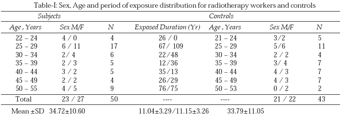
Chromosomal aberration analysis was conducted on fifty Radiotherapy workers from different hospitals of the Shahid Beheshti and Tehran Medical Universities. Chromosome alterations were analyzed, taking in to consideration the duration of exposure, age and sex. Age ranged from 22 to 55 (mean of 34.72� 10.60) years in workers and from 21 to 53(mean 33.79�11.05) years in control group. There was no significant increase in the frequency of age between workers and control groups (P = 0.337), the detail is summarized in (Table-I).
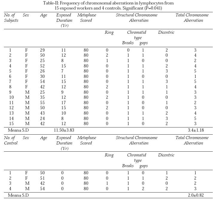
A total of 4000 metaphases or on average about 80 cells per workers and controls were analyzed. We scored more metaphases for both group bearers with chromosome aberrations. Of particular interest is that majority of dicentric is not accompanied with fragments. The incidence of structural chromosome aberrations including Dicentric, Fragments and Rings in lymphocytes from fifteen subjects (exposed group) with exposure duration from 7 to 15 years (mean frequency 11.50� 3.83) and in cells from only four control individuals is summarized in (Table-II).
There was a significant increase (P= 0.041) in the number of structural chromosomal aberration in the peripheral lymphocytes of the exposed group compare with controls. The mean values were 3.4�1.18 abnormal metaphases for workers and 2.0�0.82 for control individuals (Table-II).
Although the mean frequencies of chromosomal aberrations in the female workers (3.5�1.42) was slightly higher than in males (3.28�0.95), there was no significance differences (P=0.74) in the frequency of chromosome aberration between males and females of radiotherapy workers.DISCUSSION
There are numerous studies on the induction of chromosomal aberration by radiation; however, to our knowledge, this is the first reported study on chromosomal aberration in radiotherapy workers in Iran. Chromosomal aberration frequency provides the most reliable biological marker of dose for detecting accidental radiation exposure. Hence, the International Atomic Energy Agency (IAEA)10 in 1991 suggested that chromosomal aberration in peripheral blood lymphocytes provides a sensitive measure of the quality and quantity of radiation exposure, and chromosome aberrations have been used for biological dosiometry in human exposures to radiation.11,12 In our investigation, the majority of aberrations for both groups of subjects were dicentrics, acentrics and ring chromosomes, which is consistent with the observations of others.4-13
The yields of chromosome fragments and total aberrations were significantly higher in workers exposed for expended periods of time,14-16 and in cells from patients with various syndromes affecting DNA repair or cell cycling, like Ataxia telangiectasia.17 The data presented here and in another study indicate that low level occupational radiation exposure for relatively short periods of time, e.g., two years,18 has no significant influence on lymphocyte chromosomal aberration frequency. These data demonstrate the usefulness of chromosome aberration as a biological marker of dose and cellular damage, which is sensitive to the duration of radiation exposure in the field.19,20 In addition to structural chromosome aberration, all types of radiation affect the frequency of numerical chromosome aberrations,2 but we did not find any numerical chromosomal alteration in our investigation. Densely ionizing radiation induces severe cell cycle delay at M-phase,21 exposure to low level ionizing radiation induces chromosomal damage at G2 phase,22 and non-DNA target components of the mitotic spindle apparatus can also be involved in chromosomal aberration.23 Phosphorylation of histone H2AX at a flanking site causes DNA double-stranded breaks in cells from radiation workers.24
Neither age nor sex was a significant predictor of chromosomal aberrations in our study. Statistical analysis did not show a significant sex-age association with chromosomal aberration which is consistent with other studies.4,20,25
In conclusion, our results indicate that long-term exposure to even low doses of ionizing radiation increases the frequency of chromosome aberrations, and potentially the risk of adverse health effects. Also chromosome aberration frequency provides a reliable biological marker of dose to detect acute accidental radiation exposure. Finally, the increase in chromosome aberrations in our radiotherapy workers underscored the need of adopting measures to avoid or minimize overexposure to radiation.REFERENCES
1. Kammk R, Lobrich M. Evidence for a Lack of DNA double stranded break repairs in human cells exposed to very low X-ray doses. Proc Nate Acad Sci 2003;29:5057-62.
2. Dahle J, Kuam E. Induction of delayed mutations and chromosomal instability in fibroblasts after UVA, UVB and X-radiation. Mutat Res 2003;11:1464-69.
3. Lee YS, Lee MS, Lee JH, Kim TH, Sang JJ. Maternal or paternal exposure to radiation increases susceptibility to the induction of glutathion S-transferase positive hepatic foci in offspring rats. Cancer Lett 1998;13:231-6.
4. Rozgaj R, Suba V, Simic D. The frequency of dicentrics and acentrics and the incidence of rouge cells in radiation workers. Mutagenesis 2002;17:135-9.
5. Karthikeya B, Prabhu B, Venkatachalan P, Paul SF. Comparison of intra-and intra chromosomal aberrations in blood samples exposed to different dose rates of gamma radiations. Radiat Prot Dosimetry 2003;103:103-9.
6. Kadhim MA, Lorimore SAT, Ownsend KMS, Goodmood DT. Radiation-induced genetic instability delayed cytogenetic aberration and apoptosis in primary human bone marrow cells. Int J Radiat Biol 1995;67:287-93.
7. Lioyd DC, Edwards AA, Leonard A, Dechut GL. Chromosomal aberrations in human Lymphocytes induced in vitro by Low doses of X- rays. Int J Radiat Biol 1992;61:335- 43.
8. Diaz-Valecillo M, Fernandez R, Rojas A, ValecillosJ, Canizales J. Chromosomal alterations in workers exposed to ionizing radiation. Invest Clin 2004;45:197-201.
9. ISCN. An International System for human Cytogenetic Nomenclature. Cancer Genet. Cytogenet 1978;2133:09-14.
10. ICRP publication 60, Recommendation of International Commission on Radiological Protection, 1991.
11. Brooks AL, Khan MA, Jostes RF, Cross FT. Metaphase chromosome aberrations as a means of radiation exposed and dose. J Toxicol Environ Health 1993;40:277-88.
12. Mefferi F, Angelini S, Forti GC, Violante FS, Loi V, Mattioli S, et al. Spectrum of chromosomal aberrations in peripheral lymphocytes of hospital workers occupationally exposed to low doses of ionizing Radiation. Mutat Res 2004;547:91-9.
13. Jha AN, Sharma T. Enhanced frequency of chromosome aberration in workers occupationally exposed to diagnostic X-rays. Mut Res 1991;260:343-8.
14. Balsen AN, Ali A, Mosa HS, Hussain KO. Chromosomal aberration analysis in peripheral lymphocyte radiation workers. Mutat Res 1992;271:209-11.
15. Hayata I, Kanda R, Minamihisamatsu M, Furaunkawa M, Susaki MS. Cytogenetical dose estimation for three severely exposed patients in the JCO critically accident in Tokia Mura, J Radiat Res 2001;42:149-55.
16. Anjaria KB, Rao BS. Chromosomal aberration analysis in chronically exposed radiation workers. Envion Pathol Toxicol Oncol 2004;23:207-13.
17. Kawwata T, Ito H, George K, Wu H, Uno T, Isobe K. Radiation induced chromosome aberration in ataxia telangiectasia cells: high frequency of deletions and misrejoint detected by fluorescent in situ hybridization. Radiat Res 2003;159:597-603.
18. Norman N, Cochra S, Bass D, Roe D. Effects of age, sex and diagnostic X-ray on chromosome damage. Int J Radiat Biol 1984;46:317-21.
19. Chung HW, Eun KR, Yang JK, Sung WH. Chromosome aberrations in workers occupationally exposed to diagnostic X-ray. Mutat Res 1996;350:307-14.
20. Milacic S. Frequency of chromosomal lesions and damaged lymphocytes of workers occupationally exposed to X-rays. Health Phys 2005;88:334-9.
21. Durate M, Furusawa Y, Majima H, Kawata T, Goton E. Association between G2 phase block and repair of radiation induced chromosome fragments in human lymphocytes. Radiat Res 1999;151:670-6.
22. Pincheira P, Lopez I, Sanbueza S, Ferruz P, Navarrete M H, Santos L. G2 repair and chromosomal damage in lymphocytes from workers occupationally exposed to low level ionizing radiation. Bio Res 1999;32:297-306.
23. Sgura A, Antoccia A, Chrubini R, Tanzavella C. Chromosome non disjunction and less induced in aneuploidy. Radiat Res 2001;156:225-31.
24. Mac Phail SH, Banath JP, Yu Y, Chu E, Olive PL. Cell cycle dependent expression of phosphorylated histone H2AX: reduced expression in unirradiated but not X-irradiation G1-phase cells. Radiat Res 2003;156:759-67.
25. Zakeri F, Assaei RG. Cytogenetic monitoring of personnel working in angiocardiography laboratories in Iran. Mutat Res 2004;562:1-9.
HOME | SEARCH | CURRENT ISSUE | PAST ISSUES
Professional
Medical Publications
Room No. 522, 5th Floor, Panorama Centre
Building No. 2, P.O. Box 8766, Saddar, Karachi - Pakistan.
Phones : 5688791, 5689285 Fax : 5689860
pjms@pjms.com.pk