|
|
||||
|
Published by : PROFESSIONAL MEDICAL PUBLICATIONS |
||||
|
ISSN 1681-715X |
||||
|
||||
|
- |
||||
|
CASE REPORT |
||||
|
- |
||||
|
Volume 23 |
April - June 2007 (Part-I) |
Number 2 |
||
|
|
||||
|
|
||||
|
|
||||
|
Published by : PROFESSIONAL MEDICAL PUBLICATIONS |
||||
|
ISSN 1681-715X |
||||
|
||||
|
- |
||||
|
CASE REPORT |
||||
|
- |
||||
|
Volume 23 |
April - June 2007 (Part-I) |
Number 2 |
||
|
|
||||
|
|
||||
Diagnostic laparoscopy and resection of a rare
case of large mesenteric liposarcomaMuthukumaran Rangarajan1, Chinnusamy Palanivelu2,
Rangasamy Senthilkumar3, Madhupalayam Velusamy Madankumar4ABSTRACT
Liposarcoma arising from the mesentery of the bowel is a rare lesion. Some of the most common presenting symptoms of primary mesenteric liposarcoma is increasing abdominal girth, weight loss, abdominal pain, abdominal discomfort with meals and the presence of a freely movable abdominal mass or masses. Our patient presented with a large intra-abdominal mass. Diagnostic laparoscopy revealed tumor confined to the mesentery of the ileum. Laparoscopy was attempted, though conversion was necessary to achieve negative margins. Laparotomy was required to resect the tumour with wide margins. Myxoid and well-differentiated types of liposarcoma are by far the most common histological type. Tumor size greater than 20cm predict significantly poorer prognosis. The treatment of choice for primary mesenteric liposarcoma is surgical resection with clear margins. Radiotherapy or systemic chemotherapy has no benefit in increasing long-term survival. Laparoscopy is of limited value.
KEYWORDS: Diagnostic Laparoscopy, Mesentery Tumor, Wide excision liposarcoma.
Pak J Med Sci April 2007 Vol. 23 No. 2 267-269
1. Dr. Muthukumaran Rangarajan
Registrar in Surgical Gastroenterology.
2. Dr. Chinnusamy Palanivelu
Director
3. Dr. Rangasamy Senthilkumar,
Registrar in Surgical Gastroenterology.
4. Dr. Madhupalayam Velusamy Madankumar,
Registrar in Surgical Gastroenterology.
1-4: GEM Hospital, 45-A,
Pankaja Mill Road, Ramnathapuram,
Coimbatore � 641045,
India.
Correspondence
Dr. Muthukumaran Rangarajan
E-Mail: rangy68@gmail.com
* Received for Publication: June 19, 2006
* Accepted: October 2, 2006
INTRODUCTION
Mesenteric tumors are uncommon and are encountered in all age groups from infancy to the very elderly. They may be cystic or solid, and they may demonstrate malignant or benign clinical behavior. Primarily anecdotal references to this class of tumors have been made throughout the 20th century. In 1936, Hart provided the earliest clear description of solid mesenteric tumors.1 Published report consisted of small numbers of cases, which makes it difficult to determine the incidence of specific tumor types. Incidence ranges from one case per 200,000-350,000 population Mesenteric tumors are cystic in 40-60% of cases. Numerous reports of mesenteric lipomas in adults and children have appeared over the years, suggesting that these probably represent the most frequently encountered cause of primary mesenteric origin.2 Primary mesenteric liposarcoma is very rare, this being only the nineteenth case reported.
CASE REPORT
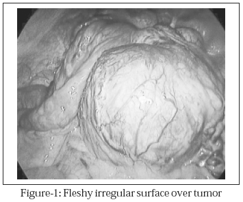
The patient was a 58-year old male who presented with a large abdominal swelling of 6 months duration. He had occasional dull-aching pain. Physical examination revealed a large mass in the central abdomen, which was well defined, had an irregular surface, hard in consistency, freely mobile and non-tender. Ultrasonogram (USG) and CT scan showed a mass measuring 14x12 cm, probably arising from the retroperitoneum. The clinical differential diagnosis was lymphoma or retroperitoneal tumor with no evidence of metastasis. The patient was planned for diagnostic laparoscopy. The mass was found completely contained within the mesentery of the ileum (Figure-1).
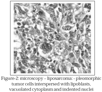
There was no local infiltration to neighboring structures. It was a vascular, fleshy tumor with irregular surface. Trial dissection was performed to assess resectability. We were able to perform dissection around the tumor with Harmonic scalpel. Since there was a possibility of tumor being malignant and risk of tumor seeding, we decided to convert to laparotomy. Also, even if it were resected laparoscopically, an incision would be necessary to deliver it out. A lower midline incision was made and the lesion was exteriorized and decompressed and the tumor was carefully delineated and dissected off the mesentery by ligating the vessels supplying it. Macroscopic tumor-free margin was 3cm. After removal of the tumor viability of the bowel in question was checked and found to be doubtful. So, 8cm of ileum was selected and end-to-end anastomosis was performed. The defect in the mesentery was closed with interrupted silk sutures. We did not choose to perform en block resection to begin with as we hoped to avoid unnecessary bowel resection, thereby avoiding morbidity. At the same time, we did not want to compromise the clearance margins. No drain was placed. The postoperative period was uneventful; liquids were started on the third postoperative day (POD), normal diet on the 5th POD and the patients was discharged on the 7th POD. There was no wound infection. Histopathological examination revealed liposarcoma, with all margin-free (Figure-2). Patient was followed up for five years, with no evidence of recurrence or metastasis.
DISCUSSION
Malignant primary mesenteric tumors are extremely uncommon. Published reports suggest that 33-50% of mesenteric masses are malignant tumors.3 These reports indicate that approximately 66% of malignant mesenteric tumors are mesenchymatous (leiomyosarcoma or liposarcoma), while the remaining is primarily lymphomas. No known etiologic or associated diseases have been reported in cases of primary mesenteric neoplasms. Intra-abdominal liposarcomas have been known to involve the retroperitoneum, jejunum, liver, stomach and esophagus. Small bowel mesentery is almost exclusively the site of involvement in the vast majority of malignant mesentery tumors. Mesenteric liposarcoma causing intestinal obstruction and peritonitis have been reported.4 A report in the year 2000 stated that only 18 cases of mesenteric liposarcoma have been reported so far.5 Clinical findings and symptoms associated with mesenteric tumors of all types are related to the presence of a mass lesion. Pain is the principal manifestation and is related to a mass effect or traction on the mesentery. Abdominal CT scans provided important information regarding size, localization of the mass and involvement of adjacent structures, as well as some tissue characteristics of the tumor.6 They can be very suggestive of lipoma, although liposarcoma cannot be completely excluded based on CT scan appearance. These lesions are of fat density and may surround mesenteric vessels and lymph nodes. MRI is sometimes used in conjunction with CT to help characterize mesenteric masses. USG is used far less frequently than CT, largely because of operator-dependent issues that can influence study consistency. Surgical treatment is the only therapy of demonstrated benefit for these tumors. The goal of surgical treatment is removal of gross disease with a margin of normal tissue. This requires resection of any involved intestine, as well as of intestine robbed of mesenteric arterial blood supply by the dissection to remove the tumor. The gross extent of the tumor must be carefully defined in order to obtain clear margins of resection. Normal intestine should be preserved to the greatest extent possible while still observing good oncologic surgical principles. Laparoscopic management has never been reported in mesenteric liposarcomas. Laparoscopy was done in this patient to confirm diagnosis and to assess resectability. The reasons for conversion have already been discussed earlier. So far, no large series reporting management and outcomes of treatment of this tumor type have ever been reported. Sato et al concluded that tumor size greater than 20cm predicted a significantly poorer prognosis.7 Published reports suggest similar biologic behavior to primary GI mesenchymal malignancies with 5-year survival rates at 20-50%.8 Postoperative adjuvant or palliative therapies are not beneficial.9 We conclude that laparoscopy has limited value in these cases and wide excision should be carried out, even if it means a large incision.
REFERENCES
1. Moyana TN. Primary mesenteric liposarcoma. Am J Gastroenterology 1988;83(1):89-92.
2. Pawel BR, de Chadarevian JP, Inniss S, Kalwinski P, Paul SR, Weintraub WH. Mesenteric pleomorphic liposarcoma in an adolescent. Arch Pathol Lab Med 1997;121:173-6.
3. Signer RD, Bregman D, Klausner S. Giant lipoma of the mesentery: Report of an unusual case and review of the literature. Am Surg 1976;42(8):595-7.
4. Manson JM. Mesenteric liposarcoma; a rare cause of intestinal obstruction. Br J Surg 1951;38(151):394-6.
5. Ciraldo A, Thomas D, Schmidt S: Giant Abdominal Liposarcoma: A Case Report. The Internet Journal of Surgery 2000;1(2).
6. Popiolek J, Szczudlinski J. Case of primary liposarcoma of the mesentery. Arch Sci Med 1982;139(4):523-9.
7. Ato T, Nishimura G, Nonomura A, Miwa K. Intra-abdominal and retroperitoneal Liposarcomas. Int Surg 1999;84:163-7.
8. Baldi A, Ganio E, Rosato L. Case of primary liposarcoma of the mesentery. Arch Sci ed 1982;139(4):523-9.
9. Orzechowski H, Wlodarczyk K. Liposarcoma of the small-intestinal mesentery as a cause of acute peritonitis. Wiad Lek 1984;37(5):385-7.
HOME | SEARCH | CURRENT ISSUE | PAST ISSUES
Professional
Medical Publications
Room No. 522, 5th Floor, Panorama Centre
Building No. 2, P.O. Box 8766, Saddar, Karachi - Pakistan.
Phones : 5688791, 5689285 Fax : 5689860
pjms@pjms.com.pk