|
|
||||
|
Published by : PROFESSIONAL MEDICAL PUBLICATIONS |
||||
|
ISSN 1681-715X |
||||
|
||||
|
- |
||||
|
CASE REPORT |
||||
|
- |
||||
|
Volume 23 |
April - June 2007 (Part-I) |
Number 2 |
||
|
|
||||
|
|
||||
|
|
||||
|
Published by : PROFESSIONAL MEDICAL PUBLICATIONS |
||||
|
ISSN 1681-715X |
||||
|
||||
|
- |
||||
|
CASE REPORT |
||||
|
- |
||||
|
Volume 23 |
April - June 2007 (Part-I) |
Number 2 |
||
|
|
||||
|
|
||||
Rhabdomyosarcoma of the larynx
Soheila Nikakhlagh1, Nader Saki2, Nepton Emad Mostofi3, Mehran Peyvasteh4ABSTRACT
Rhabdomyosarcoma of the larynx is a very unusual neoplasm and delays in diagnosis are common because the presenting symptoms are often mistaken for inflammatory or benign laryngeal disease, therefore a high index of suspicion is necessary to make diagnosis. We report a 13 years old girl with diagnosis of laryngeal Rhabdomyosarcoma. The optimum treatment of head and neck rhabdomyosarcoma has not been defined. Therapeutic modalities include an aggressive surgery without major morbidity. Radiotherapy and chemotherapy is also preferred for the treatment of rhabdomyosarcoma.
KEY WORDS: Rhabdomyosarcoma, Larynx.
Pak J Med Sci April 2007 Vol. 23 No. 2 280-282
1. Dr. Soheila Nikakhlagh
2. Dr. Nader Saki
1-2: Assistant Prof. of Otolaryngology,
Department of Otolaryngology,
3. Nepton Emad Mostofi,
Assistant Prof. of Pathology,
Department of Pathology,
Clinical and Anatomical Pathologist,
4. Dr. Mehran Peyvasteh
Department of Pediatric Surgery,
Assistant Prof. of Pediatric Surgery,
1-4: Imam Khomeini Hospital, Ahwaz, Iran.
Correspondence
Dr. Mehran Peyvasteh
E-mail: mehran.peyvasteh@gmail.com
* Received for Publication: July 1, 2006
* Accepted: November 15, 2006
INTRODUCTION
Rhabdomyosarcoma of the larynx is a very unusual neoplasm and delays in diagnosis are common.1 The presenting symptoms are often mistaken for inflammatory or benign laryngeal disease, therefore a high index of suspicion is necessary to make diagnosis.2,3
CASE REPORT
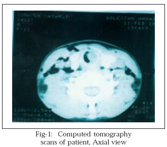
A 13 years old girl was referred to our hospital and admitted to ICU due to respiratory distress and failure to response to asthma treatment. Her main symptom was dyspnea. Physical examination showed intercostal and suprasternal notch retraction, longer inspiration than expiration, stridor, hoarsness and generalized wheezing and ronchi in both lungs. The rest of her physical exam was unremarkable. Liver and kidney function tests and chest X-ray were normal. After few hours she went into respiratory failure and was a candidate for intubation. She was unable to be intubated because of subglottic stenosis that seems to be due to a subglottic mass. After that, she was taken to the operating room and tracheostomy was done by otolaryngology service. CT scan showed a solid mass and its origin was from left subglottic wall (Fig-1).
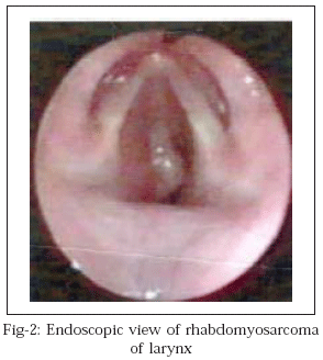
Fiberoptic laryngoscopy and bronchoscopy revealed a submucosal vegetative mass located in 1.5cm inferiorly to the vocal cords occupying 80% of laryngeal lumen and was limited to the same region (Fig-2).
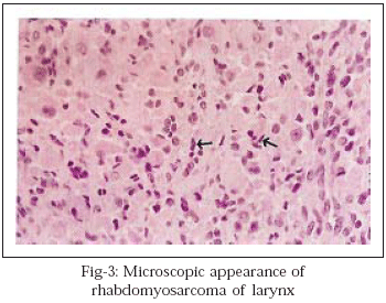
Several biopsies were performed. The histopathologic diagnosis revealed fragments of nonkeratinized squamous epithelium infiltrated by round and spindle neoplastic cells, with atypical nuclei and eosinophilic cytoplasm compatible with spindle cell Rhabdomyosarcoma (Fig-3).
Immuno histochemical study confirmed diagnosis of Rhabdomyosarcoma of larynx, botryoid variant. The patient was referred to an oncology department. She was given two courses of chemotherapy and course of radiotherapy which resulted in remission.
Discussion
Rhabdomyosarcoma is a highly malignant tumor that arises from undifferentiated mesodermal tissue.4,5 It accounts for 40% of sarcomas found in the head and neck region and is the most common sarcoma in children.6-8 The laryngeal rhabdomyosarcomas are quiet rare and might be the least common sarcoma of the larynx.9 There are four principal histologic varieties of rhabdomyosarcoma: embryonal, alveolar, pleomorphic and botryoid.8-10 The botryoid variety however is the only variant of embryonal rhabdomyosarcoma with characteristic gross appearance which was seen in our case. Most reported age for rhabdomyosarcoma are influenced by the preponderance of embryonal tumors that account for approximately two thirds of all cases and usually occur in patients younger than age 10 years and males account for approximately 55% to 70% of patients.11-13 Radical and mutilating surgical resection for the treatment of head and neck rhabdomyosarcoma has been largely replaced by the use of radiation and chemotherapy.13,14 Surgery is used to excise small readily accessible tumors or to reduce bulky tumor. This is followed by intensive treatment with other two modalities. Chemotherapy can reduce tumor size to such an extent that a large non resectable tumor may become amenable to resection.15,16 On gross pathology, rhabdomyomas are reddish-brown, lobulated, and soft.2 Histologically, these tumors are further subcategorized into adult and fetal forms according to their degree of cellular differentiation and maturity. Adult-form extracardiac rhabdomyomas are composed of closely packed round cells with peripherally located nuclei. The cells have eosinophilic vacuolated cytoplasm that is glycogen-rich. Cross-striations (similar to those in mature striated muscle) are characteristic of rhabdomyomas. Immunostaining reveals positive findings for muscle-specific actin, desmin, and myoglobin, which are markers of mature muscle cells. Mitoses are typically absent.2,6 Although isolated cases in children have been reported, adult-form rhabdomyomas present almost exclusively in patients older than 40 years. They occur more often in men by a ratio of 3:1.6 Patients may present with a palpable mass, airway obstruction, dysphagia, foreign body sensation, hoarseness, or serous otitis media caused by eustachian tube obstruction.6,7 Treatment of rhabdomyomas requires complete surgical resection. Local recurrence has been reported in more than one third of cases,6 and usually results from incomplete resection. Recurrences may present months to years after initial resection. Isolated cases of fetal- form rhabdomyoma have been associated with embryonal rhabdomyosarcoma.1 To our knowledge; there are no documented cases of malignant degeneration of adult-form rhabdomyomas. 6
REFERENCES
1. Kapadia SB, Meis JM, Frisman DM, Ellis GL, Heffner DK, Hyams VJ. Adult rhabdomyoma of the head and neck: A clinic opathologic and immunophenotypic study. Hum Pathol 1993;24:608-17.
2. Lanzkowsky P. Manual of pediatric Hematology and oncology. Third edition 2002;12:324-35.
3. David M. Pediatric Neoplasia: morphology and Biology. Lippincott-raven publishers, Philadelphia 1996.
4. Vincent T, Devita Jr SH, Rosenburg SA. Cancer: Principles and practice of oncology; 5th edition. Lippincott - raven 1997;21:257-61.
5. Batsakis JG. Tumors of the Head and Neck. Clinical and pathological considerations. Baltimore 2004;36:1647-63.
6. Kedar A, Kuten A, Joachims HZ, Arieh Y, Yudelev M. Rhabdomyosarcoma of the larynx treated by laser surgery combined with radiotherapy and chemotherapy. Med Pediatr Oncol 1983;11(4):279-80 .
7. Diehn KW, Hyams VJ, Harris AE. Rhabdoyosarcoma of the larynx. Laryngoscope 1984;94( 2pt 1 ):201-5.
8. Haerr RW, Turalba CI, El mahdi AM, Brown KL. Alveoar rhabdomyosarcoma of the larynx. Laryngoscope 1987;97( 3pt 1):339-44 .
9. Fecnandes P, Tendon DA, Tickoo SK, Rath GK. Embryonal rhabdomyosarcoma of the larynx in a child. Indian J Cancer 1988;25( 2):89-93.
10. Gross M, Gutjahr P. Therapy of rhabdomyosarcoma of the larynx. Int J Pediatr otorhinolaryngol 1988;15(1):93-7.
11. Balazs M, Egerszegi P. Laryngeal botryiod rhabdomyosarcoma in an adult pathol Res pract 1989;184(6):643-9.
12. Enzinger FM, Weiss SW. Rhabdomyoma. In: Enzinger FM, Weiss SW, eds. Soft tissue tumors, 3rd edition. St. Louis: Mosby 1995;523-36.
13. Kato MA, Flamant F, Lacombe MJ, Habrand JL, Lemerle J. Rhabdomyosarcoma of the larynx in children Med Pediatr Oncol 1991;19(2):110-4.
14. Ruske DR, Glassford N, Costello S. Laryngeal rhabdomyosarcoma in adults. J Laryngol Otol 1998;112(7):670-1.
15. Helmberger RC, Stringer SP, Mancuso AA. Rhabdomyoma of the pharyngeal musculature extending into the prestyloid parapharyngeal space. AJNR 1996;17:1115-18.
16. Mortele K, Lemmerling M, Vanzieleghem B, Moerman M, Cuvelier C, Kunnen M. Laryngeal embryonal rhabdomyosarcoma in a child. Eur Radiol 1998;8(7):1251-3.
HOME | SEARCH | CURRENT ISSUE | PAST ISSUES
Professional
Medical Publications
Room No. 522, 5th Floor, Panorama Centre
Building No. 2, P.O. Box 8766, Saddar, Karachi - Pakistan.
Phones : 5688791, 5689285 Fax : 5689860
pjms@pjms.com.pk