|
|
||||
|
Published by : PROFESSIONAL MEDICAL PUBLICATIONS |
||||
|
ISSN 1681-715X |
||||
|
||||
|
- |
||||
|
ORIGINAL ARTICLE |
||||
|
- |
||||
|
Volume 25 |
April - June 2009 (Part-I) |
Number 2 |
||
|
|
||||
|
|
||||
|
|
||||
|
Published by : PROFESSIONAL MEDICAL PUBLICATIONS |
||||
|
ISSN 1681-715X |
||||
|
||||
|
- |
||||
|
ORIGINAL ARTICLE |
||||
|
- |
||||
|
Volume 25 |
April - June 2009 (Part-I) |
Number 2 |
||
|
|
||||
|
|
||||
Study of enterococcal susceptibility patterns
isolated from clinical specimens in Tabriz, Iran
M.T. Akhi1, F. Farzaneh2, M. Oskouei3
ABSTRACT
Objectives: To identify the prevalence of enteroccoci species in clinical specimens, to determine their susceptibilities to some antibiotics for treatment, and to detect the vanA-specific 377-bp fragment from the genomic DNA of all vancomycin resistant enteroccoci (VRE).
Methodology: One hundred thirty seven isolates of enterococci species were obtained from samples of patients who were referred to microbiology laboratory of two hospitals in Tabriz from March 2001 to April 2002. After identification of enterococcal species by biochemical methods, the antibiotic susceptibility of isolates was determined by standard disk diffusion test according to NCCLS. MIC tests for vancomycin were also carried out for VRE strains by macro-dilution method. The vanA-specific 377-bp fragment was amplified from the genomic DNA of all VRE by PCR.
Results: The isolates were found to consist of E. faecalis (90.5%), E. faecium (5.84%) and Enterococcus species (3.66%). According to susceptibility data obtained, six (4.38%) out of 137 isolates were found to be VRE with MIC e"32ěg/ml. The vanA gene fragments of Enterococcus faecalis, Enterococcus faecium, Enterococcus gallinarum and Enterococcus durans, were amplified from isolates and were detected.
Conclusion: Finding of this study shows an emergence of VRE along with increased rate of resistant enterococci in Tabriz.
KEYWORDS:
Vancomycin resistant enterococci, Antibiotic resistance, VanA gene, Tabriz.Pak J Med Sci April - June 2009 Vol. 25 No. 2 211-216
How to cite this article:
Akhi MT, Farzaneh F, Oskouei M. Study of enterococcal susceptibility patterns isolated from clinical specimens in Tabriz, Iran. Pak J Med Sci 2009;25(2):211-216.
1. M.T. Akhi, Bsc., Msc., Ph.D.
2. F. Farzaneh, Bsc., Msc.
1,2: Department of Microbiology,
Faculty of Medicine,
Tabriz University of Medical Sciences,
Tabriz – Iran.
3. M. Oskouei, Bsc., Msc., Ph.D
Division of Microbiology,
Pastuer Institute of Iran,
Tehran – Iran
Correspondence
M.T. Akhi, Bsc., Msc., Ph.D.
E-mail: M_T_Akhi@yahoo.com
Akhim@tbzmed.ac.ir
* Received for Publication: September 13, 2008
* Accepted: February 2, 2009
INTRODUCTION
Enterococci are gram-positive, opportunistic bacteria that inhabit the gastrointestinal tracts of humans and many animals. Enterococci are the second most frequently reported cause of surgical wound infections and nosocomial urinary tract infections and the third most frequently reported cause of bacteremia.
1,2 Resistance to environmental conditions such as heat or desiccation allow prolonged survival and poor compliance with hand-washing procedures by health care workers results in the rapid spread of enterococci in hospitals.3,4 Moreover, strains of enterococci have acquired resistance to essentially most of the antimicrobial agents over the past three decades. Nosocomial infections with entrococci are major concern at many hospitals and have rapidly increased in many countries worldwide including Iran.5-8 Resistance of these microorganisms against the first choice therapy (aminopenicillin/aminoglycoside) required alternative treatment with the glycopeptides, vancomycin or teicoplasmid.The prevalence of vancomycin
resistance in enterococci (VRE) has dramatically increased in the last few years.9 Six types of vancomycin resistance have been reported in enterococci (VanA, VanB, VanC, VanD, VanE and VanG). The resistance to vancomycin is inducible and is encoded by the vanA gene cluster, which is carried on transposons.2 Transfer of resistance can occur via conjugative plasmids. Enterococci as reservoirs of antibiotic resistance genes, tend to transfer their resistance genes to the other bacteria among them methicillin- resistant Staphylococcus aureus.10 Monitoring the antibiotic resistance of enterococci isolated from clinical specimens is a useful tool to get information about prevalence of VRE and will be essential for controlling the spread of bacterial resistance.The aim of this study was to determine the species distribution and drug susceptibility patterns of clinical enterococcal isolates at two hospitals in Tabriz, Iran.
METHODOLOGY
Samples collection: In cross sectional study during March 2001 to April 2002, a total of 137 enterococcal isolates were recovered from clinical specimens (Urine, ascetic fluid, wound, catheter, blood, bone marrow) of patients from two clinical microbiology laboratories of Emam and Sina hospitals in Tabriz, Iran. Only one isolate per patient was included in the study. All isolates were stored in brucella glycerol broth at -20°C until tested.
11Identification of strains: Isolates were identified to the genus and species level based on the standard biochemical and microbiological methods such as: morphologic appearance on Gram-stain (gram positive cocci forming short chains), catalase negative, ability to hydrolyze esculin in the presence of bile, growth in the presence of 6.5% NaCl at 45şC, as well as commercially available kit (API 20 Strep, biomerieux, France).
11Antimicrobial susceptibility testing: Susceptibility to antimicrobial agents for all enterococci isolates was determined by the standard disk diffusion method and confirmed according to the NCCLS guidelines current at the time of study on Muller-Hinton agar incubated for 24 hr at 37şC.
12 The following antibiotic disks (BBL) were used: oxacillin (1µg), penicillin (10 units), ampicillin (10µg), amoxicillin/clavulanic acid(30µg), gentamicin (10µg), erythromycin (15µg), vancomycin (30µg), trimethoprim-sulfamethoxazole (23.75µg sulfamethoxazole, 1.25µg trimethoprim), nalidixic acid (30µg), ciprofloxacin (5µg), chloramphenicol (30µg) and nitrofurantoin (300µg). The values of minimum inhibitory concentration (MIC) of each VRE isolates for vancomycin were determined by the broth macrodilution method according to NCCLS guidelines.13 Susceptibility test results were assessed after 24-48 hr incubation at 37şC. The control strains used in this work were Staphylococcus aureus ATCC 29213 and E. faecalis ATCC 29212 for susceptibility test. Isolates with MIC of <4µg/ml were considered susceptible and with MIC of >16µg/ml were recorded as resistant.vanA gene: All VRE strains were grown for 24 hour at 37 ± 0.5°C in BHI with 5% sheep blood. DNA was extracted using sodium dodecyl sulphate- proteinase K method modified with N-cetyl-N, N, N-trimethylammonium bromide (CTAB).
14 The following oligonucleotides were used as primers for amplification of the 377-bp fragment of the vanA gene: vanA 1 (5 -TCT GCA ATA GAG ATA GCC GC-3 ) and vanA 2 (5 -GG AGT AGC TAT CCC AGC ATT-32).15(Primers were obtained from TIB MOLBIOL, Berlin, Germany). The total volume of PCR mix was 25µl, including: sterile redistilled H2O 17.05µl, 10X PCR buffer 2.5µl, dNTP mix (10mM) 0.5µl, MgCl2 (50mM) 0.75µl, forward primer (25µM) 0.5µl, reversed primer (25µM) 0.5µl, Taq DNA polymerase (5U/µl) 0.2µl, template DNA 3µl. Negative controls contained all components except template DNA.The two primers and other reagents were prepared according to the manufacturer’s recommendations. PCR reactions were performed with an automated thermal cycler (Eppendorf mastercycler gradient, Germany) with the PCR cycling conditions (initial cycle at 94şC for 4 min, 30 cycles of denaturation at 94şC for 40 sec, annealing at 57şC for 40 sec, and extension at 72 şC for 40 sec, final cycle extension at 72 şC for 7 min).
16Gel electrophoresis was performed for 60-120 min in a 1.2% agarose gel at 75 V. DNA profiles were visualized by means of ultraviolet (UV) light after ethidium bromide staining on a UV transiluminator. The gels were photographed using a gel documentation system (UVP, USA) for the analysis of bands. E. faecium, a vanA-negative strain, and vanA-positive strain E. faecium were used as reference strains.
RESULTS
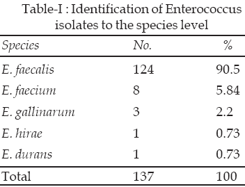
Collection of strains: A total of 137 strains were collected: 120 (87.59%) isolated from urine; five (3.65%) were from ascetic fluid; four (2.92%) were from blood; three (2.19%) were from wounds; three (2.19%) were from Catheter; and two (1.46%) were from bone marrow. Distribution of Enterococcus isolates is shown in Table-I. A total of six VRE strains isolated during the investigation period, were identified to the species level as follows: One E. faecalis, three E. faecium, one E. gallinarum and one E. durans.
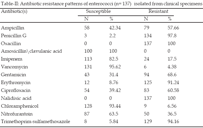
The Susceptibility data obtained in vitro for 137 isolates with 13 antibiotic substances are shown in Table-II. Multidrug-resistant isolates were found in all of isolates. Of 137 enterococci only six (4.38%) isolates were resistant to vancomycin (MICs >16Aµg/ml,). Four (66.7%) out of six isolates were from urine specimens (one E. faecalis, two E. faecium, one E. durans,). The other two VRE were isolated from blood (16.65% E. gallinarum) and catheter (16.65% E. faecium). In addition to vancomycin, all of the VRE isolates (n = 6) demonstrated resistance to a wide variety of other antimicrobial agents such as penicillin, oxacillin, ampicillin, imipenem, nalidixic acid, ciprofloxacin, trimethoprim-sulfamethoxazole, gentamicin, and erythromycin. They were susceptible to amoxicillin-clavulanic acid, chloramphenicol and nitrofurantoin.

vanA gene: The vanA-specific 377-bp gene fragments of E. faecalis, E. faecium, E. durans, and E. gallinarum, were amplified from isolates and the results obtained for some strains are shown in figure. The positive control strain E. faecium contained the typical 377-bp fragment, and the negative control strain E. faecium did not.
DISCUSSION
Resistance is known to arise where use of antimicrobial agent is high and spread of these resistance bacteria is easy. Because of the limited therapeutic options and lack of enough information and programs to control rapid spread of Enterococci species, the mortality of the enterococcal infection is on the rise, so comprehensive data concerning the susceptibility patterns of enterococcal isolates is needed to control spread of these resistant bacteria. The distribution of enterococcal species observed in this study was similar to the previous reports and E. faecalis (90.5%) was the species more frequently isolated from clinical samples.
7,17,18The resistant rate to ampicillin (57.66%) in enterococcal isolates in this study was close to the resistance rate to ampicillin in Ireland (51%).
19 However resistance rate to ampicillin reported by Mathur et al in India (66%) is higher than our result.20 Since ampicillin is the drug of choice in the treatment of enterococcal infections, the relatively high resistance of isolates in this study to ampicillin is of great concern especially in the case of endocarditis treatment. The finding that all of isolates were susceptible to amoxicillin/clavulanic acid is of great importance, and is similar to that reported in West Indies.21 Although there are reports of 46% and 8.16% enterococci resistance to amoxicillin / clavulanic acid from India.17,22 Chloramphenicol was the third most active antibiotic (93.44%) against our isolated enterococci, which is similar to findings of other studies.7,23,24The prevalence of gentamycin resistance in enterococcal isolates in present study (68.6%) was higher than that found in Ireland (60%)
19 so limiting the success of such associated antibiotic therapy. The high-level resistance to aminoglycosides is of great concern, since it eliminates synergy with cell-wall active antibiotics, a combination commonly used for the treatment of enterococcal endocarditis. The prevalence of high-level gentamicin resistance in enterococci was comparable to that reported in the antimicrobial surveillance program in Europe.9In this study 91.24% of isolates were resistant to erythromycin which is higher than what was reported from other countries such as India (85%) and Lebanon (59%).
20,23 Of the total of 137 enterococcal isolates, 83 (60.58%) were resistant to ciprofloxacin which was similar to the findings of Miskeen et al (55.78%)17 and Rudy et al25 but was much lower than the result obtained in India (88%).20.The prevalence of ciprofloxacin resistant enterococci have been reported to be 3.14%, 10% and 34% in French, Japanese and Lebanon studies respectively.23,26,27 These results indicate diverse geographic distribution of ciprofloxacin resistant enterococci. According to literature nitrofurantoin is one of the effective antibiotics on enterococci species18,25 but the results of this study showed 36.5% of resistance to nitrofurantoin, for which the possible common prescription of this drug for urinary tract infections could be the reason. The increase of resistance to nitrofurantoin in this study also corresponds to the finding of other workers in Tehran.7The concomitant resistance observed to penicillin, oxacillin, co-trimoxazole, doxycycline, nalidixic acid, and some other antibiotics confirms that resistance of enterococci to multiple antibiotics is common as it is also observed in other parts of the world.
7,21,25The reported incidence of vancomycin-resistant enterococci (VRE) isolated in hospitals throughout the world,
2,7,9,23 has been also observed in Tabriz (4.38%). This result is in agreement with the enterococcal vancomycin resistance rates in some of the European countries and Canada but less than that reported in Turkey (11.7%), Latvia (14.3%)9 and Tehran (10.6%).7Presence of vanA gene cluster, on some of our isolates can provide transfer of vancomyvin resistance via conjugative plasmids not only to enterococci species but also to other bacteria such as Staphylococcus aureus so we expect the increase of the number of VRE in the future. The resistance rate to vancomycin (4.38%) is a serious threat that necessitates using surveillance studies, infection control and monitoring of antibiotic sensitivity among hospital isolated strains.
ACKNOWLEDGEMENTS
We thank Prof. M. R. Nahaei from Tabriz University of Medical Science for his help and consultation. We also express our thanks to the Vice Chancellor for research of Tabriz University of Medical Science for financial supports.
REFERENCES
1. McCormick JK, Hirt H, Dunny Gm, Schlievert PM. Pathogenic mechanisms of enterococcal endocarditis. Curr Infect Dis 2000;2(4):315-21.
2. Courvalin P. Vancomycin- resistance in gram-positive cocci. Clin Infect Dis 2006;42 suppl 1:s25-34.
3. Zirakzadeh A, Patel R. Vancomycin resistant enterococci: colonization, infection, detection, and treatment. Mayo Clin Proc 2006;81(4):529-36.
4. Ott M, Wirick H. Vancomycin-resistant enterococci and the role of the healthcare worker. Can Oper Room Nurs J 2008;26(1):21-4,26-9.
5. Vandamme P, Vercauteren E, Lammens C, Pensart N, Leven M, Pot B et al. Survey of enterococcal susceptibility patterns in Belgium. J Clin Microbiol 1996;34(10):2572-6.
6. Kacmaz B, Aksoy A. Antimicrobial resistance of enterococci in Turkey. Int J Antimicrobial Agents 2005;25(6):535-38.
7. Feizabadi, MM, Asadi S, Aliahmadi A, Parvin M, Parastan R, Shayagh M et al. Drug resistant patterns of enterococci recovered from patients in Tehran during 2000-2003. Int J Antimicrobial Agents 2004;24(5):521-22.
8. Vilela MA, Souza SL, Palazzo IC, Ferreira JC, Morais Jr. MA, Post Darini AL et al. Identification and molecular characterization of Van A-type vancomycin-resistant Enterococcus faecalis in northeast of Brazil. Mem Inst Oswaldo Cruz 2006;101(7):715-19.
9. Schouten MA, Hoogkamp – Korstaje JA, Meis JF, Voss A. Prevalence of vancomycin resistant enterococci in Europe. Eur J Clni Microbiol Infect Dis 2000;19(11):816-22.
10. Chang S, Dawn M, Sievert MS. Infection with vancomycin- resistant Staphylococcus aureus containing the vanA resistance gene. N Engl J Med 2003;384:1342-47.
11. Domig AG, Mayer HK, Kneifel W. Methods used for the isolation, enumeration, characterization and identification of Enterococcus spp.1.Media for isolation and enumeration. Int J Food Microbiol 2003;88(2-3):147-164.
12. National Committee for Clinical Laboratory Standards: Performance standards for antimicrobial disk susceptibility tests, ed 6, Approved Standard M2-A6. Wayne, Penn, 1997, NCCLS.
13. National Committee for Clinical Laboratory Standards: Methods of dilution antimicrobial susceptibility testing for bacteria that grow aerobically, ed 4, Approved Standard M7-A4, Wayne, Penn, 1997, NCCLS.
14. Van Soolingen D, De haas P EW, Hermans PWM, Van Embden JDA.DNA fingerprinting of Mycobacterium tuberculosis. Methods Enzymol 1994;235:196-205.
15. Duka-Malen S, Molinas C, Arthur M, Courvalin P. The vanA glycopeptide resistance protein is related to D-alanyl-D-alanine ligase cell wall biosynthesis enzymes. Mol Gen Genet 1990;224(3):364-72.
16. Araj GF, Talhauk RS, Simaan CJ, Maasad MJ. Discrepancies between mecA PCR and conventional tests used for detection of methicillin resistant Staphylococcus aureus. Int J Antimicrob Agents 1999;11(1):47-52.
17. Miskeen PA, Deodhar L. Antimicrobial susceptibility pattern of Enterococcus species from urinary tract infections. J Assoc Physicians India 2002;50(3):378-81.
18. Barisic Z, Punda-Polic V. Antibiotic resistance among enterococcal strain isolated from clinical specimens. Int J Antimicrob Agents 2000;16:65-8.
19. Lavery A, Rossney, AS, Morrison D, Power A, Keane CT. Incidence and detection of multi-drug resistant enterococci in Dublin hospitals. J Med Microbiol 1997;46:150-6.
20. Mathur P, Kapil A, Chandra R, Sharma P, Das B. Antimicrobial resistance in Enterococcus faecalis at a tertiary care centre of northern India. Indian J Med Res 2003;118:25-8.
21. Orrett FA, Connors E. Enterococcal urinary tract infections: eight years experience at a regional hospital in trinidad, West Indies. Chin Med J 200;114(1):90-2.
22. Kapoor L, Randhawa VS, Deb M. Antimicrobial resistance of enterococcal blood isolates at pediatric care hospital in India. Jpn J Infect Dis 2005;58(2):101-3.
23. Zouain MG, Araj GF. Antimicrobial resistance of enterococci in Lebanon. Int J Antimicrob Agents 2001;17(3):209-13.
24. Pegues DA, Pegues CF, Hibberd PL, ford DS, Hooper DS. Emergence and dissemination of highly vancomycin- resistant vanA strain of Enterococcus faecium at a large teaching hospital. J Clin Microbiol 1997;35(6):1565-70.
25. Rudy M, Nowakowska M, Wiechula B, Zientara M, Radosz- Komoniewska H. Antibiotic susceptibility analysis of Enterococcus spp. Isolated from urine. Przegl Lek 2004;61(5):473-6.
26. Guirguitzova B, Chankova D, Zozikova B, Minkov N. Enterococci as uropathogens: frequency of isolation and sensitivity to antibacterial agents. Ann Urol (Paris) 1998;32(1):15-19.
27. Nakanishi N, Yoshida S, Wakebe H, Inoue M, Mitsuhashi S. Mechanisms of clinical resistance to flouroquinolones in Enterococcus faecalis. Antimicrob Agents Chemother 1991;35(6):1053-9.
HOME | SEARCH | CURRENT ISSUE | PAST ISSUES
Professional
Medical Publications
Room No. 522, 5th Floor, Panorama Centre
Building No. 2, P.O. Box 8766, Saddar, Karachi - Pakistan.
Phones : 5688791, 5689285 Fax : 5689860
pjms@pjms.com.pk