|
|
||||
|
Published by : PROFESSIONAL MEDICAL PUBLICATIONS |
||||
|
ISSN 1681-715X |
||||
|
||||
|
- |
||||
|
ORIGINAL ARTICLE |
||||
|
- |
||||
|
Volume 23 |
April - June 2007 (Part-II) |
Number 3 |
||
|
|
||||
|
||||
|
|
||||
|
Published by : PROFESSIONAL MEDICAL PUBLICATIONS |
||||
|
ISSN 1681-715X |
||||
|
||||
|
- |
||||
|
ORIGINAL ARTICLE |
||||
|
- |
||||
|
Volume 23 |
April - June 2007 (Part-II) |
Number 3 |
||
|
|
||||
|
||||
Expression and function of integrin associated protein
(IAP)/CD47 in human articular cartilage
Orazizadeh Mahmoud1, Salter Donald2
ABSTRACT
Background: Integrin-associated protein (IAP) /CD47, a member of immunoglobulin super family (IgSF), is expressed in a variety of cell types. In the present work, the expression pattern of IAP/CD47 in normal and osteoarthritis (OA) human articular cartilage and its role in mechanotransduction pathway following mechanical stimulation have been studied.
Methods: Using immunohistochemistry and standard western blotting the in vivo and in vitro expression pattern of CD47 in normal and OA human articular cartilage was assessed.The role of CD47 in mechanotransduction was evaluated by using an electrophysilogical method.
Results: Immuno histochemical studies showed a similar very strong expression pattern of CD47 in both normal and OA cartilage. Electrophysilogical experiments showed a role for CD47 in chondrocyte response to mechanical stimulation.
Conclusion: Very strong expression pattern of CD47 in both normal and OA chondrocyte may be a result of its critical function/s in chondrocyte homeostasis and it appears that CD47 is important in regulation of the chondrocyte response to mechanical stimulation.
KEY WORDS: CD47/IAP, Integrin, Chondrocyte, Osteoarthritis (OA).
Pak J Med Sci May - June 2007 Vol. 23 No. 3 432-437
1. Dr. Orazizadeh Mahmoud,
Department of Anatomical Sciences, Faculty of Medicine,
Ahwaz Jundi-Shapour University of Medical Sciences,
Ahwaz – IRAN.
2. Dr. Salter Donald,
University of Edinburgh,
The Queen’s Medical Research Institute,
47 Little France Crescent, Edinburgh, Scotland, U.K.
Correspondence
Dr. Orazizadeh Mahmoud,
Email: M_orazizadeh@yahoo.com
* Received for Publication: June 19, 2006
* Accepted: January 4, 2007
INTRODUCTION
Integrin-associated protein (IAP/CD47) is a 45-55 KD plasma membrane protein which is physically and functionally associated with integrin. IAP has a heavily glycosylated extra cellular IgV-like domain, a domain containing multiple membrane spanning segments and a short cytoplasmic tail. Four alternatively spliced forms have been identified which differ in the length of the cytoplasmic tail.1 IAP has a broad tissue expression and recently has been identified as a receptor for thrombospondin (TSP) family members2 and signal regulatory proteins (SIRPs).3 The roles of CD47 are being elucidated and it is coming increasingly clear that it has important roles in regulation and modulation of different integrin- mediated cell signalling.4-7
The mechanisms by which mechanical forces regulate chondrocyte function are beginning to be defined and appear to involve activation of a variety of intracellular signalling pathways, at least some of which require integrin mediated events.
It has been shown that in normal human articular chondrocytes (HAC), cyclical mechanical stimulation at 0.33Hz (2 sec on/1 sec off) results in activation of a mechanotransduction pathways such as up regulation of aggrecan gene expression,8 á5â1 integrin-dependent signalling, stretch-activated ion channels and secretion of interleukin 4 (IL-4) which, via a paracrine / autocrine signalling loop.9 In contrast, although mechanical stimulation activates á5â1 integrin- mediated signalling events, chondrocytes derived from osteoarthritic (OA) cartilage did not show alteration in levels of aggrecan or MMP3 mRNA following 0.33 Hz stimulation.10 The reasons why normal and OA chondrocyte mechanotransduction through a5b1 integrin differ is unclear.
This study was undertaken to assess the expression pattern of CD47 in normal and OA human articular chondrocytes and establish whether it has role/s in cellular responses to mechanical stimulation.MATERIALS AND METHODS
Tissue sources and assessment: Human articular cartilage was obtained, with ethical approval and patients’ consent, at operation from knee joint arthroplasty specimens and amputations for peripheral vascular in Royal Infirmary Hospital, Edinburgh, Scotland, and UK during 1998-2003. As previously described.11 The articular surface was assessed and graded macroscopically for the presence or absence of osteoarthritis using Collins grade.12 (Table-I). One set of full thickness pieces of normal and OA cartilage was snap frozen in liquid nitrogen.
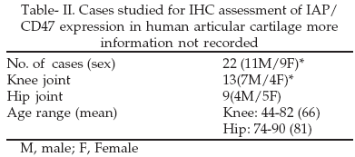
Immuno Histochemistry (IHC): Samples of normal articular cartilage (Collins grade 0).13 obtained from 1 female (age 67 years) and 7 males (median age 71 years, range 53-88) and osteoarthritic cartilage (Collins grade 1-3) obtained from 13 males (median age 71 years, range 53 - 88) were snap frozen in liquid nitrogen. 4mm sections were cut with a Brights cryostat, mounted on poly-L-lysine coated glass slides, allowed to come to room temperature and fixed with acetone for 10 min.
Sections were stained with Bric126 (1:5000) by an avidin-biotin-conjugated (ABC) (3 drops on each slide) for 30 minutes at room temperature. Slides were then visualized using diaminobenzidene (DAB) with 0.1% hydrogen peroxide (H2O2) as the peroxidase substrate for 3 min and counterstained with Harris’s hematoxyllin for 30 sec. Negative controls were provided by omitting the primary antibody. The positive immunoreactivity in different sections was graded in a range of 1-5 Table-I11 cases studied for IHC assessment of human articular cartilage are shown in Table- II.
Chondrocyte culture: Normal and OA cartilage were kept separately. Chondrocytes were isolated by sequential enzyme digestion. Briefly, cells were seeded in Iscove’s modified Dulbecco’s medium (Gibco) supplemented with 10% fetal calf serum (Sigma), 100I.U./ml penicillin (Gibco) and 100 mg/ml streptomycin (Gibco) to a final density of 2x10⁵ cells /ml (for protein extraction) and 1x10⁴ cells/ml (for electrophysiology) in 55mm plastic petri dishes (Nunc). Primary, non-confluent, 1-2 week cultures of chondrocytes were used in all experiments.
Antibodies: Mouse monoclonal antibody anti-CD47 Bric 126 (International Blood Group Reference Laboratory, Bristol, UK) for IHC and electrophysiology, and polyclonal goat anti-CD47 (Santa Cruz) for western blotting were applied.
Protein extraction and western blotting: The methods for protein extraction and western blotting used have been described previously.11 In brief, cells at rest or following mechanical stimulation were washed with ice-cold PBS containing 100µM Na3VO4 (Sigma) and lysed in situ with ice-cold lysis buffer at 40C for 15min. Lysis buffer contained 1% Igepal (Sigma), 100µM Na3VO4, and protease inhibitor cocktail tablet (Boehringer Mannheim). Supernatants were collected after centrifugation at 13,000rpm for 15 min. Concentration of protein within lysates was determined using Folin–Lowry assay method with Dynatech MR5000 and equivalent amounts loaded and separated on a 7.5% SDS-PAGE under reducing conditions. Following electrophoresis, whole cell lysates were transferred onto polyvinylidene fluoride (PVDF) membranes (Millipore Immobile-P, Sigma). Membranes were blocked overnight at 40C with 2% BSA in TBST (12.5 mM Tris/HCl, pH 7.6, 137 mM NaCl, 0.1% Tween 20). After washing with TBST, blots were incubated for one hour at room temperature with primary antibody and then HRP labelled secondary antibodies.
Electrophysiological measurements and mechanical stimulation: Membrane potentials of cells were recorded using a single electrode bridge circuit and calibrator.8 Microelectrodes with tip resistances of 40 to 60mg ohms and tip potentials of approximately 3mV were used to impale the cells. Membrane potentials of isolated cells were measured, and results were accepted if, on cell impalement, there was a rapid change in voltage to the membrane potential level that remained constant for at least 20s. The membrane potentials of 5–10 cells were measured prior to and 10 min following addition of 50µg/ml anti-CD47 Bric126. Each experiment was undertaken at least three times on cells from different donors.
Statistical analysis: The mean, standard deviation (SD) and standard error of the mean (SEM) were determined in each experiment. For statistical comparisons, the Student’s t test was applied.RESULTS
Expression of CD47 in normal human articular cartilage: Twelve sections of normal articular cartilage were obtained from 6 males (age range 62-82, mean age 71), 5 females (age range 74-90, mean age 78) and one case whose age and sex was not recorded. Normal human articular cartilage sections were isolated from 4 femoral heads, 3 females, one male (age range 74-90, mean age 79); 4 femoral condyles, 2 females, 1 male, 1 case whose age and sex was not recorded (age range 64-82, mean age 74); 3 tibial plateaux, 1 male, 1 female, one case whose age and sex was not recorded (age range 64-88, mean age 73) and 1 patella (a case whose age and sex was not recorded).
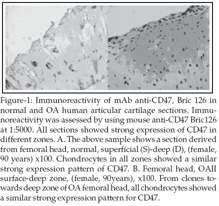
Normal articular chondrocytes in all zones exhibited a consistent pattern of CD47 expression (Figure-1A).
Chondrocytes in all zones showed very strong expression for CD47. There was no difference in the pattern of CD47 staining with respect to various zones (Table-III). The median scores for all zones of normal sections were 5. All chondrocytes in different sections from different donors showed similar staining pattern. Strong immunoreactivity of CD47 at very high dilution (1:5000) of mAb Bric 126 was observed, suggesting strong expression of CD47 in normal articular cartilage. There was no difference in immunoreactivity of sections removed from adult donors of different age or sex.
Expression of CD47/IAP in OA human articular cartilage: CD47 expression pattern was assessed in sections from 20 mild OA (7 grade I and 13 grade II) and 12 severe OA (10 grade III and 2 grade IV) of articular cartilage samples. Samples of OA articular cartilage were obtained from 11 males (age range 62-82, mean age 73), 9 females (age range 62-90, mean age 78) and 2 cases whose age and sex were not recorded. Samples of mild OA articular cartilage were obtained from 5 femoral heads, 1 male, 4 females (age range 74-90, mean 78) 8 femoral condyles, 4 males, 2 females, 2 cases whose age and sex were not recorded (age range 62-88, mean 74); 6 tibial plateaux, 3 males, 1 female, 2 cases whose age and sex were not recorded (age range 72-88, mean 75) and 1 patella, female (age 67).
Chondrocytes in all zones showed extensive membrane staining for CD47 (Figure-1B). Articular cartilage from mild OA (grades I and II) exhibited a consistent expression of CD47, which was similar in all zones of cartilage.
Strong expression of CD47 was observed in mild OA sections isolated from femoral head and various anatomical regions (femoral condyle, tibial plateau or patella) of knee joint (Table-I). The median scores for all zones of mild OA sections were 5.
Samples of severe OA articular cartilage were obtained from 6 femoral condyles, 3 males, 1 female, 2 cases whose age and sex were not recorded (age range 62-88, mean 76); 5 tibial plateaux, 3 males, 2 females (age range 62-88) and 1 patella, male (age 82). In severe OA sections (grades III and IV), chondrocytes in all zones showed strong immunoreactivity.
Strong expression of CD47 was observed in severe OA sections isolated from femoral head and various anatomical regions (femoral condoyle, tibial plateaux or patella) of knee joint. Clusters and areas of excessive surface fibrillation showed extensive anti-CD47 immunoreactivity (Figure-1B). The median scores for all zones of severe OA sections were 5 (Table-III). Different grades of OA articular cartilage did not show difference in the strong CD47 expression pattern (Table-III).
Like normal articular cartilage sections, both mild and severe OA, showed similar strong immunoreactivity of CD47 at very high dilution (1:5000) of mAb anti-CD47 Bric126, suggesting a similar expression pattern of CD47 in both normal and OA articular cartilage. CD47 positive staining did not show detectable modification in sections removed from adult donors of different age or sex.
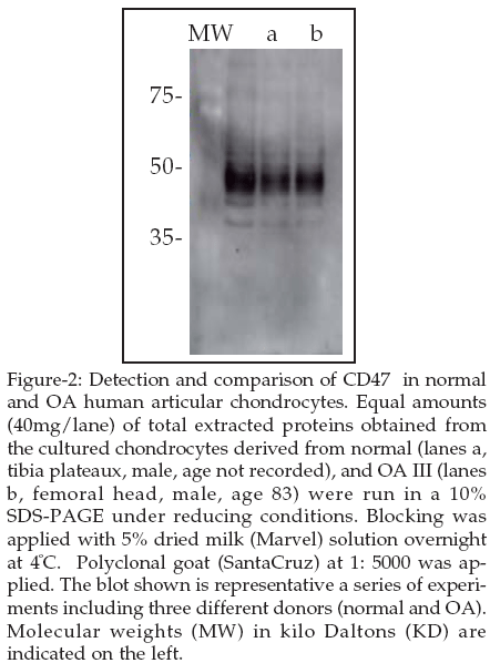
Western blotting analysis: In next step of this study, to confirm the in vivo immunohistochemical results, a series of western blotting experiments was carried out. Chondrocytes were obtained from hip, knee and ankle joints. Chondrocytes were isolated from normal articular cartilage obtained from femoral head (hip), tibial plateaux and femoral condyles (knee) of 11 males (age range 69-83, mean 75) and 2 females (age range 71-80, mean 75). OA chondrocytes were isolated from OA articular cartilage obtained from the femoral head, tibia plateaux, femoral condyles and patella of 4 males (age range 68-87, mean 77) and 2 females (age range 62-87, mean 75). Primary chondrocytes were seeded at 2x10⁵ cells/ml concentration. After a period of 10-15 days cultured chondrocytes were lysed and the proteins extracted.
There was no appreciable variation in the intensity of CD47 band between samples from normal and OA cartilage. There was no difference in results from experiments using chondrocytes removed from donors of different age and sex or from femoral head and Different areas of knee joint (femoral condyle, tibial plateaux and patella). The polyclonal goat anti-CD47 (Santa Cruz) was applied at 1: 100 dilution.
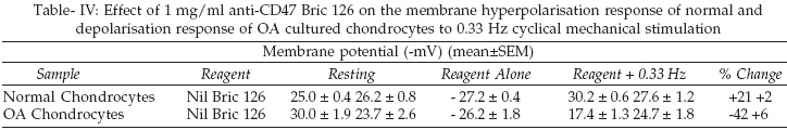
Role for CD47 in regulation of chondrocyte mechanotransduction: Following 20 min mechanical stimulation at 0.33Hz normal human articular chondrocytes show a membrane hyperpolarisation response whereas chondrocytes from osteoarthritic cartilage show a depolarisation response.The addition of 1mg/ml from anti-CD47 antibody, Bric 126, 10 minutes prior to the period of mechanical stimulation to the culture medium has no effect on the resting membrane potential of chondrocytes from either normal or osteoarthritic chondrocytes (Table-IV). Bric 126 inhibits both the hyper polarisation response of normal chondrocytes and the depolarisation response of osteoarthritic to the mechanical stimulus (Table-IV).
DISCUSSION
In this study, we have demonstrated expression of CD47 by normal and OA human articular chondrocytes with very strong expression pattern. Functional roles for CD47 in chondrocyte mechanotransduction were supported by the observations that antibodies to CD47 inhibited electrophysiological responses reproducibly seen when both normal and OA human articular chondrocytes were mechanically stimulated at 0.33Hz in an in vitro monolayer culture model system. It has previously demonstrated that á5â1 integrin has a pivotal role in human articular chondrocyte mechanotransduction potentially acting as a mechanoreceptor.8 The current study suggests that CD47 also has important roles in regulation of chondrocyte mechanotransduction, potentially through association with á5â1 integrin. CD47 has been shown to co-immunoprecipitate with a number of integrins including aVb3 (5), aIIbb3 (2á2â16 and a4b1,15 á5â1 integrin, recognised as the classical fibronectin receptor, is an important and critical candidate that could involve in the supramolecular complex formation. It has been shown that the change in membrane potential, tyrosine phosphorylation and gene expression, which follows mechanical stimulation of human articular chondrocytes, is á5â1 integrin and RGD dependent,8 although some signal transduction through CD47 is integrin-independent,16 association of CD47 with certain integrins has been found to modulate their function and CD47 has been demonstrated to functionally regulate á5â1 dependent adhesion to fibronectin,2-4 In order to find a cell surface accessory molecule(s) for á5â1 integrin, this syudy was focused on the detection and comparison in vivo expression of integrin-associated proteins (IAP)/CD47 in normal and OA human articular cartilage. It was noted that alterations in the expression or activity of one or more of the molecules involved in the signal transduction pathway could conceivably modify the response of diseased chondrocytes.
The mechanism by which CD47 influences chondrocyte mechanotransduction is unclear and may involve integrin dependent and independent pathways. Previous work in a variety of cell systems supports involvement of Gái containing heterotrimeric GTPases in CD47 regulation of integrin activity. More recently a different mechanism by which CD47 can modulate integrin affinity without intracellular signalling has been suggested. The extra cellular Ig domain of CD47 may interact with the integrin inducing change to a high affinity state presumably by modulating a structural change in the extracellular domain.
In conclusion, we have identified CD47 as a further important accessory molecule for á5â1 integrin in human articular chondrocyte mechanotransduction. Further studies are needed to evaluate the interaction between CD47 and its ligands, cross talk between it and integrins especially á5â1 integrin and its role in down stream signaling events following mechanical stimulation.ABBREVIATIONS
DAB, Diaminobenzidene; FCS, fetal calf serum; IHC, immunohistochemistry; NRS, normal rabbit serum; OA, osteoarthritis; PBS: phosphate buffered solution; RPM, revolution per minute; ECM, extracellular matrix.
ACKNOWLEDGEMENT
This work has supported by a scholarship from the Ministry of Health and Medical Education (MHME).
REFERENCES
1. Lindberg FP, Gresham HD, Schwarz E. Molecular cloning of integrin-associated protein: an immunoglobulin family member with multiple membrane-spanning domains implicated in alpha v beta 3-dependent ligand binding. J Cell Biol 1993;123:485-96.
2. Chung J, Gao AG, Frazier WA. Thrombspondin acts via integrin-associated protein to activate the platelet integrin alphaIIbbeta3. J Biol Chem 1997;272:14740-6
3. Jiang P, Lagenaur CF, Narayanan V. Integrin-associated protein is a ligand for the P84 neural adhesion molecule. J Biol Chem 1999;274:559-562.
4. Gao AG, Lindberg FP, Finn MB, et al. Integrin-associated protein is a receptor for the C-terminal domain of thrombospondin. J Biol Chem 1996;271:21-4.
5. Gao AG, Lindberg FP, Dimitry JM. Thrombospondin modulates alpha v beta 3 function through integrin-associated protein. J Cell Biol 1996;135:533-44.
6. Wang XQ, Frazier WA. The thrombospondin receptor CD47 (IAP) modulates and associates with alpha2 beta1 integrin in vascular smooth muscle cells. Mol Biol Cell 1998;9:865-74.
7. Oldenborg PA, Zheleznyak A, Fang YF. Role of CD47 as a marker of self on red blood cells. Science 2000;288(5473):2051-4 288:2051-4.
8. Wright MO, Nishida K, Bavington C. Hyperpolarisation of cultured human chondrocytes following cyclical pressure-induced strain: evidence of a role for alpha 5 beta 1 integrin as a chondrocyte mechanoreceptor. J Orthop Res 1997;15:742-47.
9. Millward-Sadler SJ, Wright MO, Lee H. Integrin-regulated secretion of interleukin 4: A novel pathway of mechanotransduction in human articular chondrocytes. J Cell Biol 1999;145:183-9.
10. .Millward-Sadler SJ, Wright MO, Davies LW. Mechanotransduction via integrins and interleukin-4 results in altered aggrecan and matrix metalloproteinase 3 gene expression in normal, but not osteoarthritic, human articular chondrocytes. Arthritis Rheum 2000;43(9):2091-9 43:2091-9.
11. Orazizadeh M, Salter DM. Differential immunohistochemical expression pattern of Galectin-3 in normal and osteoarthritic (OA) human articular cartilage. IJI 2005;2:78-86
12. Collins DH, Meachim G. Sulphate (35SO4) fixation by human articular cartilage compared in the knee and shoulder joints. Ann Rheum Dis 1961;20:117-22:117-22.
13. Lowry OH, Rosenbrough NJ, Farr AL. Protein measurement with the Folin phenol reagents. J Biol Chem 1951;193:265-75.
14. Salter DM, Millward-Sadler SJ, Nuki G. Integrin-interleukin-4 mechanotransduction pathways in human chondrocytes. Clin Orthop 2001;(391 Suppl ):S49-60S49-S60.
15. Abitorabi MA, Pachynski RK, Ferrando RE. Presentation of integrins on leukocyte microvilli: a role for the extracellular domain in determining membrane localization. J Cell Biol 1997;139:563-71.
16. Barazi HO, Li Z, Cashel JA. Regulation of integrin function by CD47 ligands. Differential effects on alpha vbeta 3 and alpha 4beta1 integrin-mediated adhesion. J Biol Chem 2002;277 (45):42859 -66 Epub 2002;277:42859-866.
HOME | SEARCH | CURRENT ISSUE | PAST ISSUES
Professional
Medical Publications
Room No. 522, 5th Floor, Panorama Centre
Building No. 2, P.O. Box 8766, Saddar, Karachi - Pakistan.
Phones : 5688791, 5689285 Fax : 5689860
pjms@pjms.com.pk