|
||||||
|
Published by : PROFESSIONAL MEDICAL PUBLICATIONS |
||||||
|
ISSN 1681-715X |
||||||
|
||||||
|
- |
||||||
|
ORIGINAL ARTICLE |
||||||
|
- |
||||||
|
Volume 23 |
January - March 2007 |
Number 1 |
||||
|
|
||||||
|
||||||
|
||||||
|
Published by : PROFESSIONAL MEDICAL PUBLICATIONS |
||||||
|
ISSN 1681-715X |
||||||
|
||||||
|
- |
||||||
|
ORIGINAL ARTICLE |
||||||
|
- |
||||||
|
Volume 23 |
January - March 2007 |
Number 1 |
||||
|
|
||||||
|
||||||
Risk factors for stroke:
A hospital based studySalma N. Khan1, Ejaz Ahmed Vohra2
ABSTRACT
Objective: Stroke is the commonest neurological cause of morbidity and mortality all over the world being the third leading cause of death. The goal of this study was to ascertain the frequency of risk factors for first ever stroke in our patients.
Patients and Methods: This prospective study included all patients of either sex, 20 to 70 years and above admitted in Dr. Ziauddin Medical University Hospital, North Nazimabad Campus, Karachi, with first ever stroke verified by CT scan brain during a period of one year.
Results: Data analysis showed that 70.1% had cerebral infarction and 29.9% cerebral hemorrhage. The mean age at presentation was 62 years and male to female ratio 1.05:1.The most frequent risk factors included hypertension 65.8%, smoking 43%, diabetes mellitus 41.3%, underlying cardiac diseases 29.1%, family history of stroke/transient ischemic attack in the first-degree relatives 26.7%, high cholesterol 25.5%, history of past transient ischemic attack 24.9% and significant extracranial carotid atherosclerosis in 18.18%. In-hospital mortality was 11.7%. At 30-day follow up 22.27% of all stroke survivors were functionally independent. This study suggested that diabetes mellitus was more and underlying cardiac diseases less frequent in our patients than in the western reported series. Cerebral hemorrhage was relatively morecommon and the mean age at presentation was lesser compared to those in the developed countries.
Conclusion: Stroke patients consume a large part of health resources all over the world so accurate information about the incidence, risk factors, management and outcome is needed for planning medico-social services besides primary and secondary stroke prevention in the community.
KEY WORDS: Stroke, Risk factors, Cerebral hemorrhage, Cerebral infarction.
Pak J Med Sci January - March 2007 Vol. 23 No.1 17-22
1. Dr. Salma N. Khan
MBBS, MD Candidate,2. Dr. Ejaz Ahmed Vohra FRCP
Professor and Head of Department,1-2: Department of Medicine,
Ziauddin Medical University Hospital,
North Nazimabad,
Karachi – Pakistan.Correspondence:
Dr. Salma N. Khan
H-2/4 Maymar Arcade
Gulshan-e-Iqbal, Block-16,
Karachi – 75300, Pakistan.
E-Mail: salmatkhan@hotmail.com* Received for Publication: March 25, 2006
* Accepted: May 26, 2006
INTRODUCTION
Stroke has been defined as acute loss of focal and at times global (applied to patients in deep coma and to those with subarachnoid hemorrhage) cerebral function; the symptoms lasting for more than 24 hours or leading to death and with no apparent cause other than vascular origin.
1 It is not a diagnosis but a clinical syndrome with numerous causes. The main types of stroke are ischemic and hemorrhagic. Defining stroke types helps in determining the most effective therapy and is clearly related to prognosis. Computed tomography or magnetic resonance imaging should be performed to confirm the type of stroke. The main goal of treatment is to maximize physical and cognitive function by limiting acute complications and facilitating rehabilitation.The studies on epidemiology of stroke are comparatively more limited in developing than developed countries. India is the only one with population-based data. The prevalence of stroke varies in different regions of India and ranges from 40 to 270 per 100,000 rural populations and is much lower from reported prevalence of 400 to 800 per 100,000 in western countries.
2 Ethnic, socio-economic and dietary factors may be responsible for this variance. Retrospective analysis of patients admitted with stroke in two hospitals of the same locality some 8 years ago in Karachi, Pakistan showed that out of the 12,454 cases 796 (6.4%) had stroke.3Epidemiologic studies of the risk factors for stroke are important for determining the origin and its prevention. In the past several decades many studies have successfully identified non-modifiable risk factors for stroke such as age, gender, race, ethnicity, heredity, and several well established modifiable risk factors also. Hypertension, atrial fibrillation, dyslipidemia, diabetes, cigarette smoking, physical inactivity, carotid stenosis, transient ischemic attack and other cardiac disorders are all potentially treatable conditions that predispose to stroke.
4Though the mortality for stroke has been on the decline still it represents the most common cause of chronic disability posing a major social and financial challenge to the community.
The objective of the present study was:-
* To ascertain the frequency of various established risk factors for stroke.
* To compare the risk factors in types of stroke (hemorrhagic vs. ischemic).
PATIENTS AND METHODS
Inclusion Criteria: All patients of either sex 20 to 70 years and above who had first ever stroke verified by CT scan brain (plain) admitted to Dr. Ziauddin Medical University Hospital, North Nazimabad Campus, Karachi, during the one-year period, from April 1
st 1997 to March 31st 1998 were included in the study.Exclusion Criteria: The following patients were excluded from the study that had
History of previous stroke*
* Subarachnoid hemorrhage
* Transient ischemic attack
* Syncopal attack
* Presumptive diagnosis of stroke with equivocal neurological deficits but no lesion on CT scan brain
* Neurological deficits secondary to epilepsy or head injury or an infective, metastatic etiology
* Pre-existing severe physical or cognitive disability.
Study Tool: After taking a verbal consent from the patients/relatives, a detailed history was taken and a thorough physical examination (including cardiovascular and neurological) was performed by the interviewer according to a self-designed stroke questionnaire. The questionnaire documented the patient’s name, age, sex, past history of transient ischemic attack and family history etc. The findings of the clinical exam were also recorded in this pre-designed form.
Stroke: The WHO definition of stroke was used. Stroke was defined as rapid onset of a new neurological deficit attributed to obstruction or rupture in the cerebral arterial system. The defined deficit had to persist for at least 24 hours unless death supervened and had to include specific localizing findings confirmed by neurological exam and by CT scan brain, with lack of evidence of an underlying non-vascular cause.
1 TIA (transient ischemic attack) was defined as rapid onset of focal neurological deficit lasting more than 30 seconds and less than 24 hours presumed to be due to cerebral ischemia and without evidence of underlying non-vascular cause.1 A CT scan brain (plain) was obtained in every patient to confirm the diagnosis and the type of stroke.The method of determining stroke type (hemorrhagic or ischemic) was similar to that used in the stroke data bank. Hemorrhagic stroke was diagnosed when intraparenchymal (within the brain substance itself) bleeding was found by CT scan and when there was no evidence on the brain image of bleeding late into an ischemic infarct. Ischemic stroke was diagnosed when a focal deficit was present and an infarct was found on CT scan or no bleeding was observed in the brain image i.e. patients with clinical features of stroke but normal CT scans were also considered to have an ischemic infarct.
5Hypertension: Patients were considered to have hypertension if they either had the diagnosis of hypertension and/or were treated for hypertension before stroke. The blood pressure was recorded after admission to the floor rather than using the emergency room measurements that were characteristically elevated. Hypertension, requiring treatment with drugs after stroke, two measurements of BP >160/95mm Hg after stroke or a single measurement of BP >180/110mm Hg were also considered to have hypertension.Patients with stroke who had transient hypertension resulting from increased intracranial pressure (Cushing reflex), who did not receive anti-hypertensive treatment and patients with BP <160/95mm Hg at the time of dismissal were not considered to have hypertension.
6-8Diabetes mellitus: Diabetes mellitus was considered present when subjects gave history of diabetes mellitus and/or were on diet/oral hypoglycemic drugs or received insulin treatment or had random blood sugar >200mg% during the hospital stay.
8Smoking: A "current smoker" was defined as a person who smoked at least one cigarette per day for the preceding three months or more or had tobacco in any form. "Ex-smoker," a person who smoked at least one cigarette per day for three months or more or had tobacco in any form at some period. "Never smoker," a person who did not meet the criteria for a current smoker or ex-smoker.
9Dyslipidemia: Dyslipidemia was defined when a patient had a diagnosis of it and/or was on diet or lipid lowering agents or had fasting blood cholesterol >200mg% in the hospital stay.
Cardiovascular causes: Patients were considered to have a cardiac abnormality when they had a self-reported history of myocardial infarction, coronary artery bypass grafting, angina or percutaneous transluminal angioplasty. The 12 lead ECG of each patient was recorded. The presence of high QRS voltage i.e. sum of S wave in V1 lead and R wave in V5 or V6 lead of 35 mm or more measures was considered evidence of left ventricular hypertrophy. ECG evidence of possible or definite myocardial ischemia i.e.1mm depression of ST segment or myocardial infarction by presence of Q/QS pattern was noted and atrial fibrillation if any was documented.
6,8Transthoracic echocardiography was done for evidence of ventricular aneurysm, mural thrombus, cardiomyopathy, hypertrophy and left ventricular hypokinesia, valvular lesions or any akinetic region was documented as a potential source of embolism in patients of cerebral infarction. A potential embolic carotid source of stroke was defined as the presence of a hemodynamically significant lesion of >70% or an ulcerated plaque seen by carotid doppler studies.
10Family History: A positive family history of stroke was considered if a patient had first degree relative (parent or sibling) who had had a stroke/TIA.
7Data was analyzed by using the software package SPSS. Qualitative variables were analyzed by finding their frequencies and percentages and Chi-square test was used to compare the risk factors in types of stroke. Quantitave variables were analyzed by calculating the mean; the standard deviation and student t test was applied to find the differences between the types of stroke. P value <0.05 was considered significant. The patients were followed up after 30-days. Those who did not turn up for follow up examination were pursued by telephonic contact.
RESULTS
Between April 1
st 1997 and March 31st 1998, 281 patients with first ever stroke admitted in Dr. Ziauddin Medical University Hospital, North Nazimabad Campus, Karachi were studied. The male: female ratio was 1.05:1. (N=281, males 144: females 137).The maximum frequency of stroke was found between the ages 51 to 70 years for both infarction and hemorrhage. The mean age was 62 years ±11.28 years. Eleven patients were younger than 41 years. Two hundred and fifty nine (92%) presented with either right or left hemiparesis/hemiplegia, 118 (42%) were unable to speak properly, 82 (29.18%) had headache, 80 (28.4%) were vomiting and 44 (15.6%) developed fits. Two hundred and five (72.95%) had Glasgow Coma Scale (GCS) between 10/15-15/15 and 76 (27%) between 3/15-9/15. Carotid bruit was audible in 45 (16%) and cardiac murmurs in 18 (6.4%).
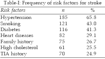
Risk factors included hypertension (HTN), smoking, diabetes mellitus (DM), underlying cardiac diseases, positive family history, high cholesterol, past transient ischemic attack (TIA) history and carotid atherosclerosis (Table-I). Mean systolic blood pressure recorded was 163±24.14mm Hg and mean diastolic blood pressure 101±44.3 mm Hg. Mean fasting blood sugar was 120±61.89mg% and mean random blood sugar 192±86.86mg%. Mean cholesterol value was 183.62±58.7mg%.
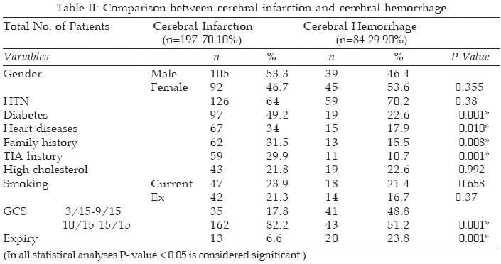
One hundred and ninety seven (70.1%) had cerebral infarction and 84 (29.9%) primary intra-cerebral hemorrhage, verified by computed tomography. In 140 (53.6%) ECGs some abnormality like left ventricular hypertrophy/hypertensive strain pattern, myocardial infarction/myocardial ischemia, atrial fibrillation was noted. It was normal in 121 (46.3%) patients. Echocardiography and carotid doppler studies were only obtained in patients with cerebral infarction.Abnormal echocardio- graphic finding as left ventricular hypertrophy, atrial enlargement, hypokinesia/akinesia, valvular lesions, ventricular aneurysm or mural clot was detected in 59 (34%) patients. In carotid doppler studies >70% stenosis/ulcerated nonstenotic plaque was observed in 28 (18.18%) patients.
Out of 281 patients 248 (88.25%) recovered while 33 (11.74%) patients died during their hospital stay; of them 20 (23.8%) had cerebral hemorrhage and 13 (6.6%) cerebral infarction. Maximum deaths were in hypertensive hemorrhagic strokes, males, 61-70 years age group with their initial Glasgow coma scale (GCS) 3/15-9/15.
Diabetes mellitus (p=0.001), heart diseases (p=0.010), family history (p=0.008), past TIA history (p=0.001) and GCS 10-15 (p=0.001) were significantly higher in patients of cerebral infarction as compared to cerebral hemorrhage while expiry (p=0.001) was significantly higher in cerebral hemorrhage. No significant difference was found in other risk factors between the two types of stroke i.e. hypertension (p=0.38), high cholesterol (p=0.992), smoking (p=0.658) and gender (0.355) (Table-II).
DISCUSSION
Although stroke mortality is declining in the west, identifying the clinical patterns and the risk factors and intervening to control or modify them remain the most important means of reducing stroke incidence.
11 Most of the local, South Asian and the far eastern studies have suggested that the proportion of intracerebral hemorrhage was significantly higher 21% to 45% than in the west 10% to 20% while cerebral infarction varied between 55% to 70.1% in the local studies and 60% to 84% in the western.6,11,12,14-24 Although some determinants of stroke, such as age, gender, race, ethnicity and heredity cannot be modified, they are risk markers. As such, they need to be considered in the patient assessments.11Increasing age is clearly the strongest determinant of the number of new cases of stroke each year. Men may be at a somewhat greater risk for stroke than women, but the difference is small. Women tend to live longer than men who die of other comorbidities; as a result, they often outnumber men in stroke prevalence figures.
11 The mean age of stroke presentation 57 to 71 years was relatively lesser here than in the west 76 to 80 years.11,13,22,25Hypertension is the most powerful and important modifiable risk factor causing a three fold greater risk of stroke than normotensive individuals.
11 In this study also hypertension was the most common risk factor similar to the other local, South Asian and western series.3,6,9,11,13-16,24,25The risk of stroke in patients with diabetes mellitus is about 4 times that found in normal individuals.
11 The frequency of diabetes mellitus was found to be higher in our population 18%-42.5% than in the western 10% to 26%.3,6,9,11,13-15,25 The estimated relative risk for stroke among smokers is 1.5 to 2.9 times that of nonsmokers. After 5–10 years, people who quit smoking reduce their risk of stroke to that of nonsmokers.11 The local studies showed somewhat similar pattern of smoking/tobacco chewing as those in the west.3,9,11,13-15,25 Cardiovascular disease is common in patients with stroke. Cardiac impairment in conjunction with hypertension further increases the risk of stroke. It increases the estimated relative risk of stroke by 2 to 4 times.11 Cardiovascular diseases were less frequent 11% to 46.5% here. Western series had much higher frequency 35%-72%.3,6,9,11,13-15,25Hypercholesterolemia and various lipoproteins fractions have been clearly associated with the severity of carotid atherosclerosis still the serum cholesterol stroke association remains an enigma.
11 It varied between 15.4% to 32% in our local series while it was 22% to 29% in the western.9,11,14,15Limitation of the study: This study done in an urban tertiary care center for a period of one year only cannot be generalized for the population at large. The most accurate measures of importance, etiological fraction and attributable risk can be estimated accurately only in large population based cohort or case control studies.
CONCLUSIONS
Stroke continues to have a great impact on public health. Stroke is frequent, recurring, and is more often disabling than fatal. The importance of preventive measures for a disease that has identifiable and modifiable risk factors must be emphasized. The reduction of morbidity and mortality among stroke patients must remain a public health priority.
REFERENCES
1. Hatano S. Control of stroke in the community, Methodological consideration and protocol of WHO stroke register. Geneva WHO 1973; document no. CVD/S/73.6 Rev.1
2. Dhamija RK, Dhamiga SB. Prevalence of stroke in rural community—an overview of Indian experience. J Assoc Physicians India 1998;46(4):351-4.
3. Vohra EA, Ahmed WU, Ali M. Etiology and prognostic factors of patients admitted for stroke. J Pak Med Assoc 2000;50(7):234-6.
4. Elkinad MS, Sacco RL. Stroke Risk Factors and Stroke Prevention. Semin Neurol 1998;18(4):429-40.
5. Davis BR, Vogt T, Frost PH. Risk factors for stroke and type of stroke in persons with isolated systolic hypertension. Stroke 1998;29:1333-40.
6. Sandercock PAG, Warlow CP, Starky IR. Predisposing factors for cerebral infarction: the Oxfordshire community stroke project. BMJ 1989;298:75-81.
7. Fiegin VL, Wiebers OD, Nikitin PY. Risk factors for ischemic stroke in a Russian community. Stroke 1998;29:34-9.
8. Caroline TM, Mackerback JP. Socioeconomic difference in stroke among Dutch elderly women. Stroke 1999;30:357-62.
9. You R, Mcneil JJ, O’malley HM. Risk factors for Lacunar Infarction Syndromes. Neurology 1995;45:1483-7.
10. Kistler JP, Ropper AH, Martin TB. Cerebrovascular diseases in Harrison’s Principles of Internal Medicine ed. 13th 1994; 2235-41.
11. Sacco RL. Risk factors and outcome for ischemic stroke, Neurology 1995;45(Suppl 1):S10-S14.
12. Banerjee TK, Mukherjee CS, Sarkhel A. Stroke in the urban population of Calcutta—an epidemiological study. Neuroepidemiology 2001;20(3):201-7.
13. Kaul S, Venketswamy P, Meena AK. Frequency, clinical features and risk factors of lacunar infarction (data from a stroke registry in South India). Neurology India 2000;48(2):1169-71.
14. Mahmood NA, Hussain T, Khan IA. Clinical spectrum of stroke in our adult population. Pak Armed Forces Med J 2003;53(1):59-67.
15. Basharat RA, Yousuf M, Iqbal J. Frequency of known risk factors for Stroke in poor patients admitted to Lahore General Hospital in 2000. Pak J Med Sci 2002;18:280-3.
16. Khan JA, Shah MA. Young Stroke - Clinical Aspects. J Coll Physicians Surg Pak 2000;10(12):461-6.
17. Khan NZ, lqbal Z. Cerebrovascular Disease, Increasing Incidence of Primary Intracerebral Hemorrhage - A Preliminary Report of 100 Cases. Pak J Neurol 1999;5(2):45-9.
18. Zhang LF, Yang J, Hong Z. Proportions of different subtypes of Stroke in China. Stroke 2003; 34:2091-95.
19. Sudlow CLM, Warlow CP. Comparable Studies of the Incidence of Stroke and its Pathological Types. Stroke 1997;28:491-9.
20. Incidence of stroke in Oxfordshire: first year’s experience of a community stroke register. Br Med J (Clin Res Ed). 1983;287(6394):713-7.
21. Bamford J, Sandercock P, Dennis M. A prospective study of acute Cerebrovascular disease in the community: The Oxfordshire community stroke project 1981-1986. J Neurol Neurosurg Psychi 1990;53(1):16-22.
22. Wolfe CDA, Rudd AG, Howard R. Incidence and case fatality rates of stroke subtypes in a multiethnic population: the South London Stroke Register. J Neurology Neurosurgery Psychi 2002;72:211-6.
23. Broderick J, Brott T, Kothari R. The Greater Cincinnati/Northern Kentucky Stroke Study. Stroke 1998;29:415-21.
24. Vohra EA, Feroz RA. Management of stroke. Specialist (Pak J Med Sci) 1990;6(2):79-85.
25. Javed MA, Ahmed M, Shahid M. Risk Factors in Stroke. Pakistan J Neurol 1998;4(1):55-8.
HOME | SEARCH | CURRENT ISSUE | PAST ISSUES
Professional
Medical Publications
Room No. 522, 5th Floor, Panorama Centre
Building No. 2, P.O. Box 8766, Saddar, Karachi - Pakistan.
Phones : 5688791, 5689285 Fax : 5689860
pjms@pjms.com.pk