|
|
||||
|
Published by : PROFESSIONAL MEDICAL PUBLICATIONS |
||||
|
ISSN 1681-715X |
||||
|
||||
|
- |
||||
|
ORIGINAL ARTICLE |
||||
|
- |
||||
|
Volume 23 |
January - March 2007 |
Number 1 |
||
|
|
||||
|
|
||||
|
|
||||
|
Published by : PROFESSIONAL MEDICAL PUBLICATIONS |
||||
|
ISSN 1681-715X |
||||
|
||||
|
- |
||||
|
ORIGINAL ARTICLE |
||||
|
- |
||||
|
Volume 23 |
January - March 2007 |
Number 1 |
||
|
|
||||
|
|
||||
Histological Study on the Effect of Tobramycin Dosage
Regimens on Renal Proximal Tubular Cells in the RatsMohamed Akram Al-Motabagani1
ABSTRACT
Objective: The present study was undertaken to show the potential nephrotoxicity of tobramycin, given in two different dosage regimens, on the proximal convoluted tubules by using the light and transmission electron microscopic techniques.
Materials and Methods: Thirty-five rats were divided into three groups: Group I served as control, Group II received tobramycin at 4mg/Kg of body weight intraperitoneally every 8 hours for ten days, and Group III received once-daily dosing of intraperitoneal injection of tobramycin at 12mg/Kg of body weight for ten days. The rats were sacrificed 3 days after the last injection. Small pieces of the right kidneys of all the animals were processed for light and electron microscopic examination.
Results: The study showed that tobramycin resulted in certain structural and ultrastructural changes in the proximal convoluted tubules. These changes included vacuolar degeneration in the epithelial cells, increased number of lysosomes with variably sized myeloid bodies, mitochondrial oedema, and loss of apical microvilli. These changes were clearly evident following multiple-daily dosing and were less obvious following once-daily dosing. Furthermore, regenerating tubular epithelial cells were evident following once-daily dosing administration.
Conclusion: The experimental tobramycin toxicity can be reduced by administering equivalent amounts of the antibiotic in a once-daily dosing as opposed to multiple-daily injections. It is hoped that this study will contribute in the selection of a more appropriate dosing regimen for tobramycin in human beings.
KEY WORDS: Tobramycin, Dosage regimens, Proximal convoluted tubules, Myeloid bodies, Regenerating cells.
Pak J Med Sci January - March 2007 Vol. 23 No.1 71-77
1. Dr. M. A. Al-Motabagani
Chairman and Head,
Department of Anatomy,
College of Medicine, King Faisal University,
P.O. Box: 2114, Dammam -31451,
Saudi Arabia.Correspondences:
Dr. Mohamed Akram Al-Motabagani,
E-Mail: drmahm@hotmail.com* Revision Received: June 21, 2006
* Revision Accepted: July 20, 2006
INTRODUCTION
Tobramycin is a member of the aminoglycosides antibiotics. The distinguishing characteristics and clinical uses of aminoglycosides have been described previously.
1 These potent antibiotics are commonly used to treat serious infections caused by many gram-negative bacilli and some gram-positive organisms. Aminoglycosides, being polar cations, are poorly absorbed from the gastrointestinal tract and hence are administered parenterally. Although they are widely used in clinical practice, serious toxicity is a major limitation to the usefulness of the aminoglycosides. Numerous toxicological studies have demonstrated the associations of nephrotoxicity, ototoxicity (vestibular and auditory), neuromuscular blockade and hypersensitivity reactions with the use of these drugs.2-9Because aminoglycosides display bactericidal, concentration-dependent killing action, there has been a long-standing debate regarding whether aminoglycosides should be administered on a once-daily dosing or multiple daily dosing regimens to achieve optimal efficacy and safety.4,8,10,11 Traditionally, the aminoglycosides have been given two to four times daily. However, recent clinical studies have suggested that less frequent, once-daily dosing, administration of aminoglycosides may achieve equal or better efficacy with less toxicity in the adults,2,4,8 children10 and neonates.12 In these clinical studies nephrotoxicity was assessed by serum concentrations analyses and immunoassay. In addition numerous studies are available describing nephrotoxicity in terms of measurements of serum creatinine and inorganic phosphate levels, levels of alkaline and acid phosphatase in kidney homogenates, detection of urinary enzymes, and sphingomyelinase and cathepsin B activities.3-12 On the other hand only few studies are available describing the morphological alterations in the renal histology after aminoglycosides administration,13-16 but did not relate the changes in the renal histology to the dosage regimens. In view of this the present work was undertaken to compare the potential nephrotoxicity of tobramycin, given in two different dosage regimen, on the proximal convoluted tubules by using the light and transmission electron microscopy.
MATERIALS AND METHODS
Thirty-five adult male Wistar albino rats were used in the present study. The weight of the rats varied from 250 to 300 grams, and their ages ranged from 9 to 11 weeks. They were fed a standard rat and mouse diet and water was supplied ad libitum. They were divided into three main groups. Group I (15 rats) served as control; five of the control rats were untreated, and the remaining ten rats were divided equally into two subgroups which received intraperitoneal injections of normal saline (NaCl, 0.9%) in the same dosage regimens as in groups II and III, respectively. Groups II and III received the same dose of intraperitoneal injections of tobramycin (Tobramycin sulfate injections, Lilly Co.) In group II (10 rats) the tobramycin was given in multiple-daily dosing in the form of a single dose (4mg/Kg of body weight) every 8 hours for ten days. Group III (10 rats) received once-daily dosing of tobramycin at 12mg/Kg of body weight for ten days. These dosage regimens were similar to those employed by previous investigators on patients undergoing treatment with tobramycin.
1,11,17The animals of all groups were sacrificed 3 days after the last injection. Prior to sacrifice the animals were anesthetized and the abdominal cavity was opened by a midline incision. The renal cortex of the right kidney of each animal was dissected, and small blocks of tissue were collected and processed for light and electron microscopy. Paraffin sections were stained with haematoxylin and eosin (H&E). Ultrathin sections were contrasted with uranyl acetate and lead citrate and examined in a Jeol JEM 100 SX transmission electron microscope operated at 80kv.
RESULTS
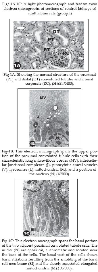
Group-I (control): The results obtained from the control group (untreated and saline-treated rats) were similar. Fig-1A, 1B and 1C are representatives of such results. Examinations of the specimens taken from the control group revealed the normal structure of the proximal convoluted tubules by both light and electron microscopy. Profiles of the proximal convoluted tubules made up the bulk of the renal cortex. The proximal convoluted tubules were lined by of a single layer of large cuboidal epithelial cells resting on a well-defined basal lamina. The nuclei were spherical and central and the cytoplasm was granular and eosinophilic (Fig-1A). Ultrastructurally, normal cellular components such as mitochondria, lysosomes, Golgi apparatus, and smooth and rough endoplasmic reticulum were present in the proximal convoluted tubules cells (Fig-1B). These cells had all the cytological features necessary for fluid and ion exchange. The apical surface had a well-developed luminal brush border consisting of numerous closely packed microvilli covered by a coat of glycocalyx. Apical canaliculi originated between the bases of the microvilli and extended into the apical cytoplasm giving rise to small pinocytotic vesicles. The vesicles coalesced to form larger vacuoles (Fig-1B). Cellular processes extended from the basolateral surfaces of each cell to interdigitate with similar processes of adjacent cells forming an extensive intricate system of interdigitating processes. Elongated mitochondria resided in the basal processes and were oriented with their long axes perpendicular to the basal lamina (Fig-1C).
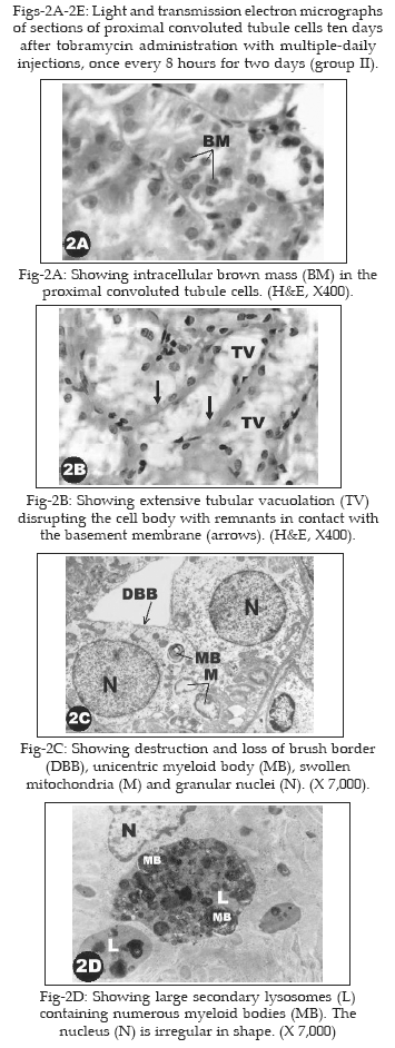
Group II (Animals treated with multiple-daily dosing of tobramycin; once every 8 hours for two days): The results obtained from the rats of group II were similar. Fig-2A, 2B, 2C and 2D are representatives of such results. In two-thirds of the proximal convoluted tubules profiles in a visual field, a brown material appeared in the form of fine discrete granules or aggregated into more prominent paranuclear masses (Fig-2A). Extensive vacuolation of the cytoplasm occurred, often disrupting the cell body so that only a cellular remnant remains in contact with the basement membrane (Fig-2B).Ultrastructurally, the brush border of the proximal convoluted tubules showed areas of complete loss and the mitochondria appeared swollen. The cytoplasm of the proximal convoluted tubule cells contained aggregates of concentrically whorled membranes (Fig-2C). The proximal convoluted tubule cells contained increased numbers of large, irregular, lysosomes with numerous myeloid bodies (Fig-2D). A few proximal convoluted tubules with minimally altered epithelium were found side by side with those having severely disrupted or sloughed epithelium. In the severely disrupted proximal convoluted tubules, there were remnants of proximal convoluted tubule cells consisting of cytoplasmic collections of disorganized, severely damaged organelles.
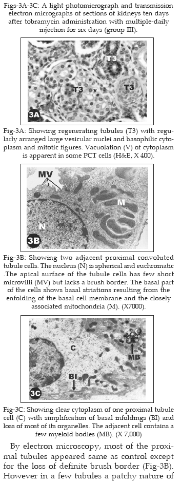
Group III (Animals treated with once-daily dosing of tobramycin for six days): The results obtained from the rats of group III were similar. Fig-3A, 3B and 3C are representatives of such results. In one-third of the proximal convoluted tubules profiles in a visual field, some of the proximal convoluted tubule cells showed vacuolation of the cytoplasm and destruction of the brush border (Fig-3A). Many proximal convoluted tubules segments were lined by regenerative epithelial cells. These cells appeared low cuboidal in shape with large vesicular nuclei and basophilic granular cytoplasm. Mitotic figures were numerous (Fig-3A).
By electron microscopy, most of the proximal tubules appeared same as control except for the loss of definite brush border (Fig-3B). However in a few tubules a patchy nature of cell injury was observed. Within the same proximal convoluted tubule, the extent of cell changes was highly variable. The cytoplasm of some cells was clear and completely disarrayed and contained few organelles, with simplifications of basal membrane infoldings. Whereas other cells in the same tubule appeared with minor changes and a few myeloid bodies (Fig-3C).
The results of the morphological changes observed with multiple-daily and once-daily dosing regimens as compared to control are summarized in Table-I.
DISCUSSION
Aminoglycosides bactericidal activity is directly related to the concentration achieved because they have a concentration-dependent killing and a concentration-dependent postantibiotic effect.
1 This enhanced activity at higher concentration, combined with the toxic side-effects, has resulted in some uncertainty with regard to the precise dosage regimen. The question of multiple-daily dosing versus once-daily dosing regimens remains in dispute. Traditionally, the total daily dose of aminoglycosides is administered as two or three equally divided doses.1 On the other hand numerous studies in a variety of clinical settings employing every commonly used aminoglycoside have favoured once-daily dosing regimen.4,8,10-12,17 In an attempt to contribute some resolution to the above dispute, the present study was undertaken with the objective of determining the effect of frequency of dosing of tobramycin on the morphology of the proximal convoluted tubules by light and electron microscopy. To achieve this objective the experimental groups of animals received equivalent total doses of tobramycin with different frequency of dosing, namely once- and multiple-daily dosing for a total duration of ten days.Examination of the renal cortex of the control group (untreated and saline-treated rats) revealed normal structural and Ultrastructural appearance. However the kidneys of the experimental (tobramycin-treated groups) revealed potential signs of nephrotoxicity with most of the damage confined to the proximal convoluted tubules. This finding is in conformity with a number of similar observations in the literature.
16,18 The simplest explanation for this phenomenon is that the proximal convoluted tubules are the largest, highly specialized segments of the nephrons and collectively make up most of the renal cortex. In addition they are the first regions of the nephrons coming in contact with drugs after being filtered by the glomeruli. In addition, accumulation of aminoglycosides in the proximal convoluted tubules cells has been attributed to the aminoglycosides-concentrating capacity of these cells.18Nephrotoxicity due to aminoglycosides has been ascribed
to endocytosis and sequestration of the aminoglycosides to lysosomes, formation of myeloid bodies, and phospholipidosis. Rupture of the lysosomal membrane leads to release of acid hydrolases with subsequent necrotic cell death.19,20 However, an alternative mechanism mediating aminoglycosides cellular toxicity has also been identified in the cell-surface, namely G protein-coupled Ca2+ (polyvalent cation)–sensing receptor (CaR). It has been suggested that the presence of the CaR in PCT, in conjunction with the ability of the aminoglycosides to activate the receptor might be involved in the process of cellular toxicity induced by the aminoglycosides.16 The current study has shown that tobramycin administration resulted in certain morphologic changes in proximal convoluted tubules. These changes included vacuolar degeneration in the epithelial cells, increased number of lysosomes with variably sized myeloid bodies, mitochondrial oedema, and loss of apical microvilli (Table-I). It is interesting to note that these changes were clearly evident following multiple-daily injections whereas following once-daily administration the morphological changes were less obvious. This may be regarded as indicative of the marked effect of frequency of dosing on tobramycin-induced morphological changes because tobramycin was introduced at equivalent total dosage. This mean that the tobramycin-induced tubular damage increases as the interval of administration is reduced from 24 to 8 hours. From these findings it may be concluded that tobramycin-induced toxicity is directly proportional to the frequency of dosing. It is possible that the intracellular accumulation of tobramycin18 resulting from activation of CaR receptors16 and binding of the tobramycin to the acidic phospholipids21 in the brush border membrane of proximal convoluted tubule cells with subsequent receptor-mediated endocytosis22 were markedly enhanced following multiple-daily as compared to once-daily injections of equivalent amounts of the antibiotic tobramycin. It is also possible that the renal clearance and elimination of tobramycin was higher following once-daily dosing.1 Moreover, it has been known that an increase in fluid influx results in cellular changes such as vacuolar degeneration, mitochondrial swelling and loss of microvilli, and that these changes are reversible.23 Thus it may be inferred that the fluid influx in the proximal tubular epithelial cells was markedly enhanced following multiple-daily as compared to once-daily dosing regimens.In the present work, the regeneration of tubular epithelium was evident following administration of the tobramycin at once-daily injections; many mitotic figures were observed in the regenerating tubules. The regenerating epithelial cells had large vesicular nuclei and basophilic cytoplasm. Electron microscopy revealed that these cells were squamoid in shape with few apical microvilli. The nucleus was flat with large prominent nucleolus, and the cytoplasm contained many mitochondria. Similar patterns of tubular regeneration have been described previously.
24,25 The appearance of the regenerating cells following administration of aminoglycosides was attributed to the morphologic and metabolic immaturity of these cells which may reflect a degree of protection from the toxic effects of tobramycin.26 The regenerating cells probably arise from the relatively unaffected residual epithelial cells that can be found in areas of tubular damage.27 It has also been reported that damage to epithelial cells in the proximal convoluted tubules may in some manner act as a stimulus to regeneration by release of cellular substances.28Since renal injury was mild following tobramycin administration at once-daily dosing regimen, coupled with the appearance of regenerating epithelial cells, it may be concluded that the experimental tobramycin toxicity can be reduced by administering equivalent amounts of the antibiotic in a once-daily dosing as opposed to multiple-daily dosing injections, which is important for the discussion of the most appropriate dosing regimen for aminoglycosides in humans.
REFERENCES
1. Chambers HF. The Aminoglycosides. Goodman & Gilman’s The Pharmacological Basis of Therapeutics, Hardman JG, Limbird LE& Gilman AG ( Eds), McGraw-Hill, New York 2002;1219-38.
2. Freeman CD, Nicolau DP, Belliveau PP, Nightingale CH. Once-daily dosing of aminoglycosides: review and recommendations for clinical practice. J Antimicrob Chemother 1997;39:677-86.
3. Buijk SE, Mouton JW, Gyssens IC, Verbrugh HA, Bruining HA. Experience with a once-daily dosing of aminoglycosides in critically ill patients. Intensive Care Med 2002;28:936-42.
4. Cohen E, Dadashev A, Drucker M, Samra Z, Rubinstein E, Garty M. Once-daily versus twice-daily intravenous administration of vancomycin for infections in hospitalized patients. J Antimicrob Chemother 2002;49:155-60.
5. Curtis JM, Sternhagen V, Batt D. Acute renal failure after placement of tobramycin-impregnated bone cement in an infected total knee arthroplasty. Pharmacotherapy 2005;25:876-80.
6. Hoffmann IM, Rubin BK, Iskandar SS, Schechter MS, Nagaraj SK, Bitzan MM. Acute renal failure in cystic fibrosis: Association with inhaled tobramycin therapy. Pediatr Pulmonol 2002;34:375-7.
7. Lerner AM, Cone LA, Jansen W. Randomized, controlled trial of the comparative efficacy, auditory toxicity, and nephrotoxicity of tobramycin and netilmicin. Lancet 1983;1:1123-6.
8. Olsen KM, Rudis MI, Rebuck JA, Hara J, Gelmont D, Mehdian R, et al. Effect of once-daily dosing vs. multiple daily dosing of tobramycin on enzyme markers of nephrotoxicity. Crit Care Med 2004;32:1678-82.
9. Paterson DL, Robson JM, Wagener MM. Risk factors for toxicity in elderly patients given aminoglycosides once daily. J Gen Intern Med 1998;13:735-9.
10. Contopoulos-Ioannidis DG, Giotis ND, Baliatsa DV, Ioannidis JP. Extended-interval aminoglycoside administration for children: a meta-analysis Pediatrics 2004;114:111-8.
11. Sung L, Dupuis LL, Bliss B, Taddio A, Abdoll M, Allen U, et al. Randomized controlled trial of once- versus thrice-daily tobramycin in febrile neutropenic children undergoing stem cell transplantation. J Natl Cancer Inst 2003;95:1869-77.
12. Thureen PJ, Reiter PD, Gresores A, Stolpman NM, Kawato K, Hall DM. Once- versus twice-daily gentamicin dosing in neonatese"34 weeks gestation: cost-effectiveness analyses. Pediatrics 1999;103:594-8.
13. Mantovani A, Macri C, Stazi AV, Ricciardi C, Guastadisegni C, Maranghi F. Tobramycin-induced changes in the renal histology of fetal and newborn Sprague-Dawley rats. Teratog Carcinog Mutagen 1992;12:19-30.
14. Mandal AK, Bennett WM. Transmission electron microscopy of urinary sediment in the assessment of aminoglycoside nephrotoxicity in the rat. Nephron 1988;49:67-73.
15. Toubeau G, Maldague P, Laurent G, Vaamonde CA, Tulkens PM, Heuson-Stiennon JA. Morphological alterations in distal & collecting tubules of the rat renal cortex after aminoglycoside administration at low doses. Virch-ows Arch Cell Pathol Incl Mol Pathol 1986;51:475-85.
16. Ward DT, McLarnon SJ, Riccardi D. Aminoglycosides increase intracellular calcium levels and ERK activity in proximal tubular OK cells expressing the extracellular calcium-sensing receptor. J Am Soc Nephrol 2002;13:1481-9.
17. Sanchez-Alcaraz A, Vargas A, Quintana MB, Rocher A, Querol JM, Poveda JL, et al. Therapeutic drug monitoring of tobramycin: once-daily versus twice-daily dosage schedules. J Clin Pharm Ther 1998;23:367-73.
18. Kaloyanides GJ, Pastoriza-Munzo E. Aminoglycoside nephrotoxicity. Kid Intern 1980;18:571-82.
19. Giuliano, RA, Paulus, GJ, Verpooten GA, Pattyn UM, Pollet DE, Nouwen EJ, et al. Recovery of cortical phospholipidosis and necrosis after acute gentamicin loading in rats. Kidney Int 1984;26:838–47.
20. Kaloyanides GJ. Aminoglycoside nephrotoxicity. Diseases of the Kidney, Schrier RW, Gottschalk CW. ( Eds), Little Brown, London. 1993;1131-64.
21. Molitoris B A, Meyer C, Dahl R, Geerdes A. Mechanism of ischemia-enhanced aminoglycoside binding and uptake by proximal tubule cells. Am J Physiol Renal Physiol 1993;264:907-16.
22. Nagai J, Takano M. Molecular aspects of renal handling of aminoglycosides and strategies for preventing the nephrotoxicity. Drug metabolism and pharmacokinetics 2004;19:159-70.
23. Mitchell RN, Cotran RS. Cell injury, death and adaptation, ‘Basic Pathology’(sixth edition), Kumar V, Cotran RS & Robbins SL (Eds), W.B.Saunders Co., Philadelphia and London 1997;4-24.
24. Luft FC, Rankin LI, Sloan RS, Yum MN. Recovery from Aminoglycoside nephrotoxicity with continued drug administration. Antimicrob Agents Chemother 1978;14:284-7.
25. Hottendorf GH, Gordon LL. Comparative low dose nephrotoxicities of gentamicin, Tobramycin, and amikacin. Antimicrob Agents Chemother 1980;18:176-81.
26. Houghton DC, Hartnett M, Campbell-Boswell M, Porter G, Bennett W. A Light and Electron Microscopic Analysis of Gentamicin Nephrotoxicity in Rats. Am J Pathol 1976;82:589-612.
27. Houghton DC, Plamp III CE, Defehr JM, Bennett W, Porter G, Gilbert D. Gentamicin and Tobramycin Nephrotoxicity. Am J Pathol 1978;93:137-52.
28. Cuppage FE, Tate A. Repair of the nephron following injury with mercuric chloride. Am J Pathol 1967;51:405-29.
HOME | SEARCH | CURRENT ISSUE | PAST ISSUES
Professional
Medical Publications
Room No. 522, 5th Floor, Panorama Centre
Building No. 2, P.O. Box 8766, Saddar, Karachi - Pakistan.
Phones : 5688791, 5689285 Fax : 5689860
pjms@pjms.com.pk