|
|
||||
|
Published by : PROFESSIONAL MEDICAL PUBLICATIONS |
||||
|
ISSN 1681-715X |
||||
|
||||
|
- |
||||
|
ORIGINAL ARTICLE |
||||
|
- |
||||
|
Volume 24 |
January - March 2008 |
Number 1 |
||
|
|
||||
|
|
||||
|
|
||||
|
Published by : PROFESSIONAL MEDICAL PUBLICATIONS |
||||
|
ISSN 1681-715X |
||||
|
||||
|
- |
||||
|
ORIGINAL ARTICLE |
||||
|
- |
||||
|
Volume 24 |
January - March 2008 |
Number 1 |
||
|
|
||||
|
|
||||
Vitro antifungal activity ofIn
Doughari JH1, Nuya SP2
ABSTRACT
Objective: To evaluate the antifungal activity of root extracts of Deterium microcarpum against some pathogenic fungi.
Methodology: Soxhlet extraction of the active principles of the plant was carried out using petroleum ether, methanol and 70% methanol in water and tested for antifungal activity against some pathogenic fungi namely; Trichophyton mentagrophytes, Aspergillus niger, Penicillium digitatum, Fusarium moxysporum, Candida albicans and Cryptococcus neoformans at varying concentrations of 100, 200, 500, 1000, and 2000 µg/ml using the filter paper disc diffusion method. The minimum inhibitory concentration (MIC) and the minimum fungicidal concentration (MFC) was also determined at extract concentration range of 65-1000 µg/ml at room temperature and 37 şC for C. albicans.
Results: The extracts were active against all the fungi tested at all concentrations of the extract used with Candida albicans showing the least susceptibility. The MIC and MFC values for the extracts ranged between 50-1000 µg/ml.
Conclusion: Antifungal substances can be sourced from Deterium microcarpum for possible development of antifungal drugs for the treatment of fungal infections such as candidiasis, cryptococcosis and skin infections.
KEY WORDS: Deterim microcarpum, Antifungal activity, Minimum inhibitory concentration, Minimum fungicidal concentration.
Pak J Med Sci January - March 2008 Vol. 24 No. 1 91-95
1. Doughari JH,
2. Nuya SP,
1-2: Department of Microbiology,
School of Pure and Applied Sciences,
Federal University of Technology,
P.M.B. 2076 Yola 640002,
Adamawa State,
Nigeria.
Correspondence
Dr. Doughari JH,
E-mail: jameshamuel@yahoo.com
* Received for Publication: July 9, 2007
* Revision Received: October 5, 2007
* Revision Accepted: October 8, 2007
INTRODUCTION
The study of resistance to antifungal agents has lagged behind that of antibacterial resistance.
1 Mycotic infectious rate increased significantly in association with several changes in medical practice, including more widespread use of therapies that depress the immune system, the frequent and often indiscriminate use of broad-spectrum antibacterial agents, the common use of indwelling intravenous devices, and the advent of chronic immunosuppressive viral infections such as AIDS.2 For nearly 30 years, amphotericin B, which is known to cause significant nephrotoxicity, was the sole drug available to control serious fungal infections. The approval of the imidazoles and the triazoles in late 1980s and early 1990s were major advances in the ability to safely and effectively treat local and systemic fungal infections. The high safety profile of triazoles, in particular fluconazole, has led to their extensive usage. Fluconazole has been used to treat in excess of 16 million patients, including over 300,000 AIDS patients, in the United States alone since the launch of this drug.1 Concomitant with this widespread use, there have been increasing reports of antifungal resistance.3 These developments and the associated increase in fungal infections4 intensified the search for new, safer, and more efficacious agents to combat serious fungal infections. Deterium microcarpum (Caesalpiniodeae Subfamily Leguminosae) is a plant with dense rounded crown shaped leaves and disc-shaped fruits (4cm diameter) with a stem covered with a brown brittle skin that grows up to 10-25m high. The seed is enclosed within an edible sweet greenish coloured pulp mixed with tangle fibre. The Nyandang and Lunguda people of Adamawa State, Nigeria call it gwogwo and takalaka respectively, while the Hausa (Northern Nigeria) call it taura. It is commonly used in the treatment of skin and scalp infections, and diarrhea associated with ‘greenish’ stooling in children. However, despite the local usage of Deterium microcarpum as a medicinal plant, there is a dearth of published medicinal research information on the plant which informs our choice of the plant for evaluation of the antifungal activity.METHODOLOGY
Laboratory grades of Petroleum ether (BDH) methanol (BDH) and acetone (BDH), and potato dextrose agar (PDA) (Difco; Detroit, Michigan) used for this work were obtained from the Microbiology Laboratory, Federal University of Technology, Yola, Nigeria.
The plant material was collected from the outskirts of the Federal University of Technology Yola, Nigeria and identified by Mr D.F. Jatau of the Department of Forestry and Wildlife Management, School of Agriculture and Agricultural Technology, Federal University of Technology Yola, Nigeria. The roots were chopped into pieces using clean sterile knife, shade-dried (30-32şC), coarsely grounded using pestle and mortar and then reduced to powder using an electric blender (National, MX-795 N) and then stored in well stoppered bottles until use.
The Microorganisms used included standardized fungal strains of Aspergillus niger, Trichophyton mentagrophytes obtained from the Microbiology Laboratory of the National Veterinary Research Institute (NVRI) Vom, Nigeria, clinical isolates of Penicillium digitatum, Fusarium oxysporum, obtained from the Microbiology Laboratory of the Specialist Hospital Yola, Nigeria and Laboratory isolates of Cryptococcus neoformans and Candida albicans obtained from the Microbiology Laboratory of the Department of Microbiology, Federal University of Technology Yola, Nigeria. All the isolates were subcultured unto fresh Potato dextrose agar slants and stored at room temperature.
Preparation of crude extracts: Four hundred grammes (400g) of the powdered plant material in each batch was exhaustively extracted by soxhlet extraction method using 1000ml of either Petroleum ether, (A), or Methanol, (B), or 70% v/v Methanol in H
2O, (C). The solvent used in each batch was recovered under pressure until dry extracts were obtained and then stored separately in amber colored bottles labeled as A, B and C. The percent yield was 35.8% (petroleum ether, 22.5% (methanol) and 41.7% for methanol water.Determination of phytochemical constituents: The freshly prepared extract was subjected to standard phytochemical analyses for different constituents such as tannins, alkaloids, flavonoids, anthraquinones, glycosides, saponins and phenols as earlier described.
5Assay of antifungal activity: The paper disc diffusion method
6 was used with slight modification. 10 ml Potato Dextrose Agar (PDA) (Difco) was dispensed into Petri dishes and allowed to solidify. Spores were recovered by gently swabbing the surface of the culture plates with a sterile cotton swab and the swab dipped in 5 ml sterile saline containing 0.1% Tween 80 in a test tube to suspend the spores. The suspended spores were diluted serially and adjusted to initial inoculum of 4 × 10⁴ spores/ml by counting using a counting chamber. A micropipette was used to introduce 0.1ml of the spore suspensions (adjusted to 4 × 10⁴ conodia/ml) on to the agar plate, and spread with glass rod spreader under sterile conditions. Sterilized discs (6mm, Whatman No 1) were prepared by soaking in different concentrations of the extracts (100, 200, 500, 1000, and 2000µg/ml) for 6 h in bijou bottles. The discs were then removed and allowed to dry in a sterile Petri dish, then stored in screw capped bottles for further use. To assay for antifungal activity, five of the discs impregnated with different concentrations of the extract were placed on a fungal spore seeded plate with the help of sterile forceps. For standard antifungal agents, sterilised discs (6mm, Whatman No 1) were also prepared by soaking in 100µg/ml concentrations of nystatin and fuchsin for 6 h in bijou bottles. The discs were then removed and allowed to dry in a sterile Petri dish and were also placed on the fungal spore seeded plate with the help of sterile forceps as positive control, while discs soaked in sterilised distilled water only without extracts were used as negative control. Three replicates were produced for each fungus. Culture plates containing C. albicans were incubated at 37 °C for 24 h while other culture plates containing the rest of the fungi were incubated at room temperature (32-35 °C) for 48-72 h. Antifungal activity was determined by measurement of the zone of inhibition (mm) around the discs after the period of incubation.Determination of minimum Inhibitory Concentration (MIC) and Minimum Fungicidal Concentration (MFC): The MIC was determined using the agar dilution method
7 with slight modifications. Varying concentrations (1000, 700, 500.0, 400.0, 300.0, 200.0, 100.0, 50.0, 25.0 and 12.5µg/ml), of the extracts were prepared and incorporated separately into respective PDA plates. All the plates were incubated at 25 şC for 48 h and inhibition of growth was noted. The MICs were recorded after 48 hour. To determine the MFC, plates which did not show any growth after 48 h from the MIC determination were further incubated for 72 hour and after incubation, the concentration at which no visible growth was seen was noted as the MFC.RESULTS
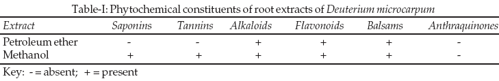
Qualitative analysis for phytochemical constituents from this study revealed the presence of saponins, tannins, alkaloids, flavonoids and balsams (Table-I).
The study revealed that Deterium microcarpum possessed significant activity against some pathogenic fungi with the methanol extracts demonstrating the highest activities of 22 and 20mm diameter of zone of inhibition against T. mentagrophytes and P. digitatum respectively, followed by those against A niger, Cr. neoformans and F oxysporum (16mm zone diameter of inhibition in each case) at 2000µg/ml (Table-II).
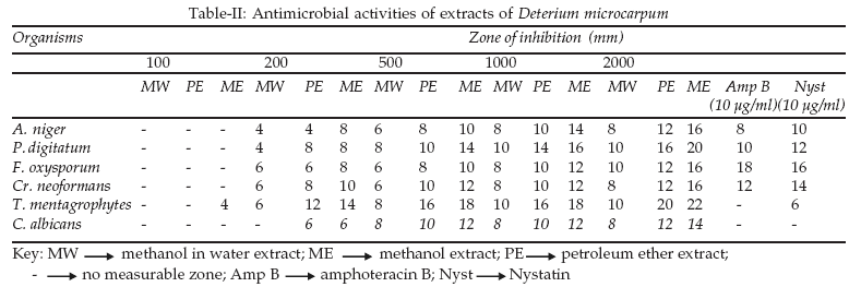
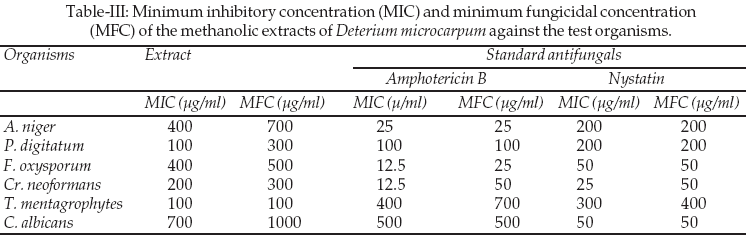
The least activity (14mm zone diameter of inhibition) was demonstrated against C. albicans at 2000 µg/ml. Methanol water extracts demonstrated the least activity. For the standard antifungal agents the highest activity (18 mm zone diameter of inhibition) was demonstrated by amphotericin B compared to that of nystatin (16 mm zone diameter of inhibition) against F. oxysporum. The least activities of 8 and 6 mm zone diameter of inhibition were demonstrated by amphotericin B and nystatin against A. niger and T. mentagrophytes respectively. C. albicans was resistant to both the two antifungal agents. Table-III shows the results of determination of minimum inhibitory concentration (MIC) and minimum fungicidal concentration (MFC) the extracts of D. microcarpum and the standard antifungal agents amphotericin B and nystatin. Results showed that, MIC and MFC values for the extracts ranged between 100-200 µg/ml with the least MIC and MFC values of 100µg/ml each demonstrated against P. digitatum and T. mentagrophytes respectively, while the highest values of 700µg/ml (MIC) and 1000µg/ml (MFC) against C. albicans and 400µg/ml (MIC) and 700µg/ml (MFC) against A. niger. For the standard antifungal agents, MIC and MBC values ranged between 12.5-700µg/ml for amphotericin B and 25-400µg/ml for nystatin with amphotericin demonstrating the least MIC value (12.5µg/ml) against F. oxysporum and Cr. neoformans compared to nystatin (25µg/ml) against Cr. neoformans.
DISCUSSION
The exhibition of antifungal activity has been attributed to the presence of plant bioactive compounds (Table-I) which are employed as natural defense mechanisms against pathogenic bacteria, fungi, viruses and pests.
8 All the fungi including A. niger were reasonably susceptible to the extracts (Table). This outcome is contrary to earlier reports 9-12 in which the organism was resistant to all the extracts and antifungal agents tested.9-12 Amphotericin B (AMB) is considered the "gold standard" for treatment of these infections. AMB is however associated with a number of severe and sometimes life threatening side effects including fever, chills and nephrotoxicity. Other treatment regimens include the ‘azole’ antifungal drugs, of which fluconazole and itraconazole are the most widely used. There is however lack of efficacy of and the emergence of resistance to itraconazole and also, limited efficacy of itraconazole against pulmonary aspergillosis. This highlights the need for new antifungal agents with potent and broad spectrum fungicidal activities for the effective management of these infections.13 The extracts were active against agents of invasive fungal infections which are important causes of death in severely immunocompromised patients and since the early 1990s invasive aspergillosis has become predominant.14 Resistance demonstrated by these fungi in previous investigations encouraged the use of drug combinations (amphotericin B (AMB) and rifampin (RIF)) to investigate antifungal activity.12 The fungi used in this study are causative agents of various mycotic infections; C. albicans (Candidiasis), Cr. neoformans (cryptococcosis), A. niger (systemic mycosis-aspergillosis) and T. mentagrophytes (ringworm). The higher MBC values to the corresponding MIC values from our data (Table-III) suggest that the fungal agents could be fungistatic at lower concentrations. Novel antifungal substances can be sourced from Deterium microcarpum for possible development of antifungal drugs for the treatment of systemic and superficial mycotic infections. Further studies on a wide range of bacteria and fungi should however be carried out.REFERENCES
1. Mahmoud AG, Louis BR. Antifungal Agents: Mode of Action, Mechanisms of Resistance, and Correlation of These Mechanisms with Bacterial Resistance. Clin Microbiol Rev 1999;12(4):501-17.
2. Joseph M, Debbie TAD, Paul EV. Use of Turbidimetric Growth Curves for Early Determination of Antifungal Drug Resistance of filamentous fungi. Clin Microbiol Rev 2003;41(10):4718-25.
3. Rex JH, Rinaldi M, Pfaller M. Resistance of Candida species to fluconazole. Antimicrob Agents Chemother 1995;39:1-8.
4. Beck-Sagué CM, Jarvis WR. National Nosocomial Infections Surveillance System. Secular trends in the epidemiology of nosocomial fungal infections in the United States, 1980-1990. J Infect Dis 1993;167:1247-51.
5. Jigna P, Nehal K, Sumitra C. Evaluation of antibacterial and phytochemical analysis of Bauhinia variegate L. bark. African J Biomed Res 2006;9(1):53-6.
6. Junaid SA, Olabode AO, Onwuliri FC, Okworiu AEJ, Agina SE. The antimicrobial properties of Ocimum gratissimum extracts on some selected bacterial gastrointestinal isolates. African J Biotech 2006;5(22):2315-21.
7. Doughari JH. Antimicrobial Activity of Tamarindus indica Linn. Tropical J Pharm Res 2006;5(2):597-603.
8. El-Mahmood AM, Ameh, JM. The in vitro antibacterial activity of Parkia biglobosa (Jacq.) root bark extract against some microorganisms associated with urinary tract infections African J Biotech 2007;6(11):57-60.
9. Lauer BA, Reuer LB, Schroter GPJ. Susceptibility of Aspergillus to 5-fluorocytosine and amphotericin B alone and in combination. J Antimicrob Chemother 1979;4:375-80.
10. Odds FC. Interactions among amphotericin B, 5-fluorocytosine, ketoconazole, and miconazole against pathogenic fungi in vitro. Antimicrob Agents Chemother. 1982;22:763-70.
11. Denning DW, Hanson LH, Perlman AM, Stevens DA. In vitro susceptibility and synergy studies of Aspergillus species to conventional and new agents. Diagn Microbiol Infect Dis 1992;15:21-4.
12. Majid Z, Mohamed RN. In vitro activities of amphotericin-B in combination with rifampin against Aspergillus species. Pak J Med Sci 2007;23(3):323-5.
13. Salama SM, Atwal H, Gandhi A, Simon J, Poglod M, Montaseri H, ET AL. In vitro and In vivo activities of Syn2836, Syn2869, Syn2903, and Syn2921: New series of triazole antifungal agents, Antimicrob Agents Chemother 2001;45:2420-6.
14. Singh N. Trends in the epidemiology of opportunistic fungal infections: Redisposing factors and the impact of antimicrobial use practices. Clin Infect Dis 2001;33:1692-6.
HOME | SEARCH | CURRENT ISSUE | PAST ISSUES
Professional
Medical Publications
Room No. 522, 5th Floor, Panorama Centre
Building No. 2, P.O. Box 8766, Saddar, Karachi - Pakistan.
Phones : 5688791, 5689285 Fax : 5689860
pjms@