|
|
||||
|
Published by : PROFESSIONAL MEDICAL PUBLICATIONS |
||||
|
ISSN 1681-715X |
||||
|
||||
|
- |
||||
|
CASE REPORT |
||||
|
- |
||||
|
Volume 25 |
January - March 2009 |
Number 1 |
||
|
|
||||
|
|
||||
|
|
||||
|
Published by : PROFESSIONAL MEDICAL PUBLICATIONS |
||||
|
ISSN 1681-715X |
||||
|
||||
|
- |
||||
|
CASE REPORT |
||||
|
- |
||||
|
Volume 25 |
January - March 2009 |
Number 1 |
||
|
|
||||
|
|
||||
Echinococcal tension pneumothorax
in a pregnant womanHasan Ekim1, Meral Ekim2
SUMMARY
Pulmonary hydatid cyst in pregnancy is a very rare pathology and its diagnosis and treatment is still a complex of problem. We report a rare case of ruptured giant pulmonary hydatid cyst presenting with tension pneumothorax during pregnancy. According to our knowledge this is the first report of such a case. A 21-year old pregnant woman was admitted to our hospital with complaints of left-sided chest pain, cyanosis and dyspnea. Chest radiograph showed tension pneumothorax, mediastinal shift, and tracheal displacement. Echocardiography revealed perforated hydatid cyst adjacent to pericardium. She was taken to the operating room immediately. During operation, a giant perforated hydatid cyst (12x10cm) was found, outside the pericardium displacing and compressing the left lower lobe. Histopathological examination confirmed the diagnosis. Approximately 5 months later she had a spontaneous vaginal delivery. Both the patient and her baby were healthy. Perforated pulmonary hydatid cyst should be kept in mind in the differential diagnosis of tension pneumothorax in a pregnant woman and surgical intervention should be performed promptly.
KEY WORDS: Pregnancy, Tension Pneumothorax, Hydatidosis.
Pak J Med Sci January - March 2009 Vol. 25 No. 1 159-161
How to cite this article:
Ekim H, Ekim M. Echinococcal tension pneumothorax in a pregnant woman. Pak J Med Sci 2009;25(1):159-161.
1. Hasan EKIM, MD,
Department of Cardiovascular Surgery,
2. Meral EKIM, MD,
Department of Biochemistry,
1,2: Yüzüncü Yil University,
Van, Turkey.
Correspondence
Dr. Hasan EKIM
E Mail: drhasanekim@yahoo.com
* Received for Publication: September 19, 2008
* Revision Received: September 23, 2008
* 2nd Revision Received: December 29, 2008
* Final Revision Accepted: December 30, 2008
INTRODUCTION
Hydatidosis is a zoonotic infestation that has been known since the time of Hippocrates. Although it is endemic in South America, Asia, Australia, and in the Mediterranean region, including Turkey; owing to the increase in worldwide travel and immigration, cardiotho-racic surgeons everywhere must be aware of this disease.
1 Tension pneumothorax is a serious but rare complication of hydatid disease. This complication can be fatal and should be kept in mind in the clinical course of hydatidosis.2 Pulmonary hydatid cyst in pregnancy is a very rare pathology and its diagnosis and treatment is still a complex of problem.3 We report a rare case of ruptured giant pulmonary hydatid cyst presenting with tension pneumothorax during pregnancy. According to our knowledge this is the first report of such a case.CASE REPORT
A 21-year old pregnant woman, whose family lives in a rural area and breeds animals, was admitted to our hospital with complaints of left-sided chest pain, cyanosis and dyspnea. Her physical examination revealed severe respiratory distress with a respiratory rate of 37 beats per minute (bpm), tachycardia of 120/bpm, blood pressure of 80/60 mm Hg, and decrease breath sounds at the left hemithorax.
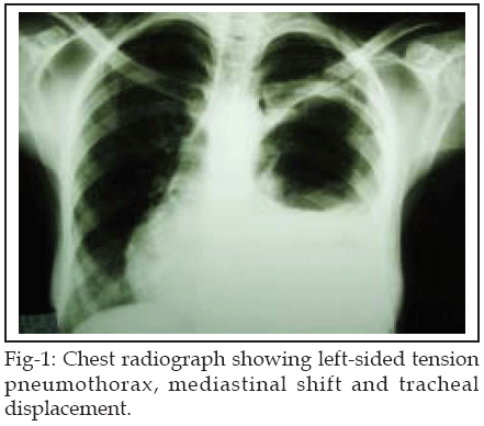
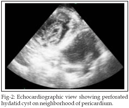
Chest radiograph taken on admission showed tension pneumothorax, mediastinal shift, and tracheal displacement (Fig-1). Echocardiography showed perforated hydatid cyst adjacent to pericardium (Fig-2). Electrocardiogram showed sinus tachycardia. The ultrasound scan confirmed a single 16-week viable fetus. Renal and hepatic function tests were normal. Indirect hemagglutination test for echinococcus was negative.
She was taken operating room immediately. Tube thoracostomy was placed, where there was a considerable air leak and some fluid came out. Left anterolateral thoracotomy was performed through the 5
th intercostals space. During operation, a giant perforated hydatid cyst (12x10cm) was found, outside the pericardium displacing and compressing the left lower lobe. After aspiration of pleural space, germinal membrane was removed as a whole with a ring forceps. The pericystic cavity was irrigated with hypertonic saline solution. Small bronchial openings were found using saline and with application of positive intrapulmonary pressure, air escaping through any bronchial opening was easily seen with the formation of air bubbles. We closed them separately with 4-0 Vicryl in a figure of eight fashion. And we confirmed the integrity of our closure by asking anesthetist to give a positive pressure. Then the residual cavity was obliterated with separate purse-string sutures that were placed into the cavity from the deepest level to the surface (capitonnage). Opposing surfaces were then sutured in an approximate face-to-face fashion to eliminate dead space. Postoperative recovery was uneventful and she was discharged on the seventeenth postoperative day. Histopathological examination demonstrated multiple endocysts with viable germinative epithelium and scoleces, confirming the diagnosis of hydatidosis.
The patient completed a normal pregnancy. Approximately five months later she had a spontaneous vaginal delivery. Both the patient and her baby were healthy. Albendazole treatment (10mg/kg) was prescribed after delivery. No recurrence was seen during one year follow-up.
DISCUSSION
Hydatid disease usually occurs in people who are living in endemic areas and who are in contact with farming animals, such as sheep and dogs. However, it has been suggested that the ingestion of raw or uncooked vegetables, such as salads contaminated with echinacoccus cysts, can act as a source of infestation.
4Pulmonary hydatid cysts are asymptomatic until they reach a large size and become complicated.
2 A sudden rise in the intrapulmonary pressure is the usual precipitating factor in rupture of the cyst: the cause could be a trivial one such as coughing or sneezing, though sometimes it may follow an increase in intraabdominal pressure as in pregnancy. However rupture may occur spontaneously without any predisposing factor.5 The occurrence of tension pneumothorax because of the rupture of the hydatid cyst is a rare complication. The signs of tension pneumothorax include marked dyspnea, cyanosis, tachycardia and hypotension. The trachea is usually deviated to the contralateral side and the affected side of the chest demonstrates increased resonance to percussion and decreased air entry and breath sounds.6 In this case, chest radiograph and echocardiography were of great value in diagnosing the perforated pulmonary hydatid cyst adjacent to pericardium.The parenchymal cavity and the bronchopleural fistula together are subject to check-valve mechanism and cause tension pneumothorax.
2 The collapsed or crumpled wall of a ruptured cyst simply acts as a curtain or check valve, admitting air into the pleura during insipiration, then falling back and closing the opening or outlet during expiration, and thus allowing air to enter but not to escape from the pleural cavity. A positive pressure will continuously build up inside the pleural cavity which will ultimately shift the mediastinal structures to the opposite side, interfering with the hemodynamics of respiratory and cardiovascular systems. These changes are responsible for the clinical picture usually seen in tension pneumothorax.5It is possible that the decrease in cellular immunity that accompanies pregnancy might be responsible for the progression of the hydatid disease, which in turn could have played a role in precipitation of rupture.
7 This same mechanism may be responsible for the cyst rupture in our patient. Serological tests are less reliable in pregnancy because of the usual immunological charges.8 Therefore, the decrease in cellular immunity that accompanies pregnancy might result in negative serology in this case.Pregnancy and hydatidosis occur occasionally together and there is no consensus about treatment. However, the treatment of hydatidosis in pregnancy is mainly surgery. But the timing of surgery is controversial and the surgery may be difficult.
3 Although some authors have reported that adjuvant albendazole treatment might be teratogenic in the first trimester but can be prescribed safely in the second and third trimesters,9 others have shown that albendazole has teratogenic effects in animals, so they have not suggested albendazole treatment during pregnancy.3,10 We do not suggest albendazole treatment before delivery. Therefore, albendazole treatment was prescribed after delivery in this case.CONCLUSION
Perforated pulmonary hydatid cyst should be kept in mind in the differential diagnosis of tension pneumothorax in a pregnant woman and surgical intervention should be performed promptly. Adjuvant albendazole treatment is essential to avoid recurrence. But, this therapy should be used after delivery.
REFERENCES
1. Kavukçu S, Kilic D, Tokat AO. Parenchyma-preserving surgery in the management of pulmonary hydatid cysts. J Invest Surg 2006;19:61-8.
2. Kurkcuoglu IC, Karaoglanoglu N. Tension pneumothorax associated with hydatid cyst rupture. J Thorac Imaging 2002;17:78-80.
3. Polat Ç, Sývacý R, Koţar MN. Recurrent hepatic hydatid cyst in a pregnant woman. Med Sci Monit 2007;13:CS27-CS29.
4. Kothari P, Aly HS, Makker HK. A solitary pulmonary cystic mass resulting from hydatid disease mimicking a pericardial cyst. Hosp Med 2001;62:640-1.
5. Bakir F, Al-Omeri MM. Echinococcal tension pneumothorax. Thorax 1969;24:547-56.
6. Hollins GW, Beattie T, Harper I. Tension pneumothorax: report of two cases presenting with acute abdominal symptoms. J Accid Emerg Med 1993;10:43-4.
7. Hijazi MH, Al-Ansari MA. Pulmonary hydatid cyst in a pregnant patient causing acute respiratory failure. Ann Thorac Med 2007;2:66-8.
8. Rahman MS, Rahman J, Lysikiewicz A. Obstetric and gynaecological presentations of hydatid disease. BR J Obstet Gynaecol 1982;89:665-70.
9. Can D, Oztekin O, Oztekin O. Hepatic and splenic hydatid cyst during pregnancy. Arch Gynecol Obstet 2003;268:239-40.
10. Monterola C, Espinoza R, Munoz S. Abdominal echinococcosis during pregnancy: A clinical aspects and management of a series of cases in chile. Tropical Doctor 2004;34:321-3.
HOME | SEARCH | CURRENT ISSUE | PAST ISSUES
Professional
Medical Publications
Room No. 522, 5th Floor, Panorama Centre
Building No. 2, P.O. Box 8766, Saddar, Karachi - Pakistan.
Phones : 5688791, 5689285 Fax : 5689860
pjms@pjms.com.pk