|
|
||||||
|
Published by : PROFESSIONAL MEDICAL PUBLICATIONS |
||||||
|
ISSN 1681-715X |
||||||
|
||||||
|
- |
||||||
|
ORIGINAL ARTICLE |
||||||
|
- |
||||||
|
Volume 24 |
July - September 2008 |
Number 4 |
||||
|
|
||||||
|
||||||
|
|
||||||
|
Published by : PROFESSIONAL MEDICAL PUBLICATIONS |
||||||
|
ISSN 1681-715X |
||||||
|
||||||
|
- |
||||||
|
ORIGINAL ARTICLE |
||||||
|
- |
||||||
|
Volume 24 |
July - September 2008 |
Number 4 |
||||
|
|
||||||
|
||||||
Endoscopic assessment of effacement of balloon
waist during pneumatic dilatation of primary
achalasia cardia under topical anesthesia
Qurratulain Hyder1, Rakhshanda Rashid2,
Syed Fazle Hadi3, Tabassum Qamar4
ABSTRACT
Objective: Balloon dilatation of Primary Achalasia Cardia (PAC) is usually performed under antegrade endoscopic guidance, with conscious sedation. The main goals of this prospective study were to assess the safety and efficacy of pneumatic dilatation without conscious sedation and to determine the endoscopic signs of effacement of the balloon"waist".
Methodology: Pneumatic dilatation was successfully performed as outdoor procedure without conscious sedation in patients (n= 25; mean age 42.56 years) with endoscopic and radiologic diagnosis of PAC.
Results: Immediate relief of symptoms was observed in 23 (92%) cases. Effacement of the balloon "waist" under endoscopic vision was appreciated in all cases in the present study. Common complications of pneumatic dilatation were chest pain in all (100%) subjects and mild local bleeding in 17 (68%) patients. There was no cancellation of procedure. Re-dilatation was required in 2 (8%) cases. The duration of follow-up was from 6 weeks to 23 months.
Conclusions: Pneumatic dilatation of PAC can be safely performed as same day procedure, without conscious sedation. Obliteration of the balloon "waist" can be readily determined by antegrade "endoscopic assessment of stretch on the lower oesophageal sphincter (EASL)".
KEY WORDS: Primary Achalasia Cardia, Pneumatic Dilatation, Endoscopic assessment, Oesophageal sphincter.
Pak J Med Sci July - September 2008 Vol. 24 No. 4 491-496
How to cite this article:
Hyder Q, Rashid R, Hadi SF, Qamar T. Endoscopic assessment of effacement of balloon waist during pneumatic dilatation of primary achalasia cardia under topical anesthesia. Pak J Med Sci 2008;24(4):491-6.
1. Qurratulain Hyder,
MBBS, FCPS
2. Rakhshanda Rashid MBBS, FCPS
3. Syed Fazle Hadi, MBBS, FRCP
4. Tabassum Qamar, MBBS, FCPS
1-4: Department of Gastroenterology,
Pakistan Institute of Medical Sciences,
G/8-3, Islamabad, Pakistan.
Correspondence
Dr. Qurratulain Hyder,
Assistant Professor, Dept. of Gastroenterology,
Pakistan Institute of Medical Sciences, G/8-3,
Islamabad, Pakistan.
E-mail: haiderkh@ucmail.uc.edu
qhyder2194@hotmail.com
* Recevied for Publication: March 5, 2008
* Revision Received: July 25, 2008
* Revision Accepted: July 28, 2008
INTRODUCTION
Primary Achalasia (PAC) is reported as an uncommon cause of dysphagia in outdoor patients.
1 Upper gastrointestinal endoscopy and barium study may be normal in early cases and in the non-classic variant. Endoscopic treatment provides the most encouraging results in PAC. Pneumatic dilatation and injection of botulinum toxin (Botox) in the lower oesophageal sphincter (LES) are now universally preferred to surgical myotomy.2-5 Modified indigenous balloons have been successfully used.6 Pneumatic dilatation was carried out under fluoroscopic guidance until recently7 but the endoscopic method has become more popular due to convenience and lack of radiation exposure.8,9 However, the outcome of endoscopic method depends on multiple factors.10Newer therapeutic techniques are likely to emerge with clinical application of translumenal surgery.
11 The present study shows that pneumatic dilatation of PAC can be safely performed without conscious sedation and fluoroscopy. As a same day procedure, this cost effective approach eliminates the adverse effects of sedation and minimizes the need for indoor observation or assisted ventilation. The endoscopic assessment of obliteration of the balloon "waist" by "EASL" has not been previously described. We also share our experience about the correlation of subjective relief with radiological improvement and the role of manometry in the management of PAC.METHODOLOGY
The present study includes 25 patients, who received endoscopic treatment (pneumatic dilatation) for PAC between October, 2004 and September, 2006. There were fourteen (56%) male and eleven (44%) female patients from 16 to 71 years of age (mean age: 42.56 years). One female patient had 28 week gestation. The diagnosis of PAC was made by upper gastrointestinal endoscopy and barium swallow examination. Patients with oesophageal strictures were excluded from the study. Informed consent was obtained for pneumatic dilatation without conscious sedation.
The patients were preoperatively reassured that the procedure-related chest pain was imminent though bearable in most instances. The equipment included a video gastroscope (2.8mm accessory channel: Pentax), Rigiflex balloon (3 cm: Microvasive), guide wire and the pressure gauge. The patients were advised nil per oral (NPO) for 6-12 h before dilatation. After topical pharyngeal anaesthesia with Xylocaine (2%), the endoscope was introduced in left lateral position. Maximum oesophageal clearance was achieved directly with the gastroscope. The guide wire was placed in the stomach through the accessory channel. The endoscope was retrieved and Rigiflex balloon was advanced over the guide wire. The gastroscope was re-introduced. The deflated balloon was negotiated across the LES under direct vision, leaving less than proximal 1/3 of its length above the sphincter. The endoscope was positioned at approximately 5cm proximal to the balloon for an antegrade view of the LES. The balloon catheter was held at the mouth guard to prevent its distal migration during air insufflation. The patient was warned about chest pain immediately before inflating the pneumatic balloon. Two successive sessions of dilatation for 30-45 seconds each, were performed with 10-15 psi inflation pressure. The LES zone was constantly observed with pre-positioned gastroscope for adequacy of dilatation, oesophageal tear and hemorrhage. The pneumatic balloon was then deflated and retrieved alongwith the guide wire. The gastroscope was removed after thorough inspection of the dilated LES. Clear fluids were permitted orally after 4-6 hours and gradually increased if the patient remained asymptomatic. A barium oesophagogram was obtained within 24 hours to exclude oesophageal perforation. Re-dilatation was planned if the symptoms persisted or there was only partial subjective relief.
RESULTS
There was immediate symptomatic improvement in twenty three (92%) of our patients after pneumatic dilatation of PAC. During follow-up period from six weeks to 23 months, re-dilatation was required in two (8%) cases within two months due to recurrence of dysphagia (Figure-1A).

The commonest complications were chest pain in all (100%) subjects and mild bleeding at the LES in 17 (68%) patients (Figure-1B). One patient experienced transient neurogenic symptoms, necessitating postponement of procedure. Another patient had minor oesophageal trauma due to accidental displacement of the guide wire. Both these patients were observed indoor for 24 hours. There was no oesophageal perforation or clinically significant hemorrhage. There was no mortality. No patient reported with gastroesophageal reflux during the follow-up period.
DISCUSSION
Achalasia cardia is the third leading cause of dysphagia in our hospital population, preceded by malignant and benign oesophageal strictures. The primary variant appears to be the most prevalent. None of our patients showed abnormal work-up, suggestive of secondary achalasia cardia. Retained oesophageal contents constitute an important clue to the endoscopic diagnosis of PAC.
12 The liquid and food debris in the oesophagus predisposes these patients to pulmonary aspiration and respiratory embarrassment during endoscopic treatment. Attempting oesophageal clearance with a nasogastric tube often induces regurgitation as the tube repeatedly curls within the oesophageal lumen due to obstruction at the LES level. We, therefore, performed endoscopic suction of the liquid component and by-passed or pushed the semi-solid contents distally during advancement of pneumatic balloon across the LES under direct vision. Our experience appears particularly safe in high-risk groups, like the elderly persons, morbid obesity, co-morbidity and in pregnant females.13,14 The procedure was well tolerated without conscious sedation. Because sedation is a continuum, the dividing line between conscious sedation and deep sedation remains indistinct. The response of an individual patient thus remains unpredictable. Prior sedation in pneumatic dilatation of PAC is more likely to enhance the risk of aspiration by central respiratory depression and blunting of cough reflex.15 Lying position further promotes gravitation of oesophageal contents into the airways. Sedation would have worsened hypotension in our patient, who experienced neurogenic symptoms during the first session of dilatation. It may be likewise detrimental during shock due to serious haemorrhage during dilatation.14,16 Sedation was also hazardous for one pregnant female in our study.17 In the absence of sedation, the need for resuscitation/assisted ventilation and the cost of indoor observation and are significantly reduced.18 We believe that a conscious patient is adequately responsive to guide the physician about intensity of chest pain vis-a�vis force of dilatation being applied during the procedure. The dilatation was thus temporarily interrupted if so desired by our patients. Conscious sedation is possibly avoided on similar grounds during dilatation of oesophageal strictures and screening endoscopy.16 Hence, we find it more prudent to eliminate the hazards of sedation by compromising for imminent though transient and bearable chest pain during pneumatic dilatation of PAC. We successfully used 3cm Rigiflex balloon both for initial and second dilatation. With constant parameters of pneumatic dilatation in the present study, we were able to achieve symptomatic relief and radiologic improvement in twenty three (92%) cases without significant morbidity. These results are comparable with several studies.6,19-21 Endoscopic guidance is superior to fluoroscopy as it obviates exposure to radiation, particularly in younger individuals and in pregnant females.22 Antegrade endoscopic view provides an excellent opportunity for real-time inspection of a fully stretched LES. We could instantly exclude oesophageal tear, laceration and active bleeding during endoscopic intervention. Leaving 1/3 length of pneumatic balloon above the LES permits larger intraluminal space for endoscopic vision.
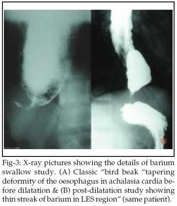
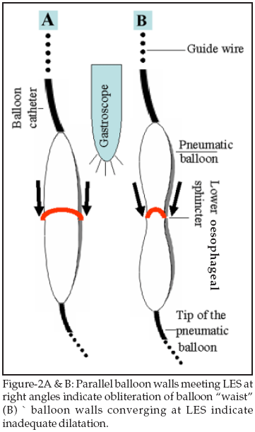
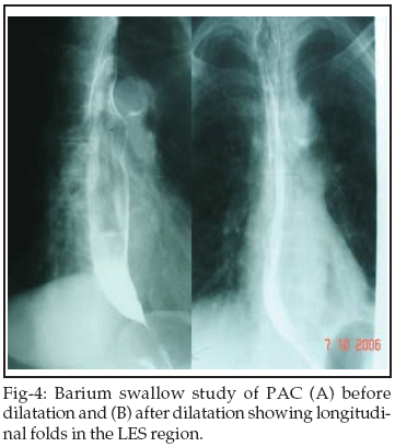
The balloon catheter needs to be held by the technician at the mouth guard to prevent its distal migration during air insufflation. We suggest "EASL" as a safer and an equally effective alternative to fluoroscopic assessment of dilatation of the LES. On looking at the LES from above, if the balloon walls run parallel and meet the stretched LES at right angles, the balloon "waist" is surely obliterated and the dilatation force is usually effective (Figure-2A): the dilatation of LES is likely to be inadequate if the balloon walls appear to converge at the LES (Figure-2B). "EASL" permits full distension of pneumatic balloon even without the use of a pressure guage. If parallel balloon walls continue to meet the stretched LES at right angles, further expansion may result in balloon rupture. Thus "EASL" can impart near-accurate information about effacement of the balloon "waist" and adequacy of LES dilatation. When combined with history of subjective improvement, "EASL" can surpass the need for serial LES manometry and radionuclide transit studies.23 However, we support the indication of an early post-operative contrast study to exclude oesophageal leak or perforation.24 Dilatation trauma to the LES results in muscle spasm in a zone with high resting pressures. Therefore, post-treatment flow of barium across the LES may not be apparent within 24 hours in spite of significant subjective relief.25 An early re-dilatation should be withheld in such cases. We encountered this situation in three (12%) patients. In our opinion, replacement of the classic "bird beak" deformity (Figure-3A) by a thin streak of barium (Figure-3B) or appearance of longitudinal folds within the LES region (Figure-4) indicate significant radiological improvement.
Radiography and endoscopy provide conclusive diagnostic information in majority of the patients with PAC. The use of oesophageal manometry, therefore, appears less justified before the initial dilatation. It seems more rational at re-dilatation when it would rule out symptom recurrence due to oesophageal dysmotility and/or LES hypertonicity. Re-dilatation was required in two (8%) of our patients. Successful re-dilatation in these subjects without the use of manometry indicates that the failure of initial intervention was primarily because of LES hypertension. Subjective and radiologic improvement in the remaining twenty three (92%) patients conforms with our observation that manometry has a limited role in the management of PAC. Some patients with long standing PAC become sitophobic. Therefore, a gradual rather than abrupt return to regular diet is better tolerated by these individuals.
CONCLUSION
Pneumatic dilatation of PAC can be safely performed under endoscopic guidance as an outdoor procedure. It is an equally effective intervention even without conscious sedation. The adequacy of LES dilatation can be readily appreciated by "EASL". Immediate post-operative radiologic assessment may be misleading due to spasm of the LES. The role of oesophageal manometry in PAC is limited. It appears to have little relevance on initial dilatation.
REFERENCES
1. Hirano I. Management of achalasia. Therapy of digestive disorders. M Michael Wolfe, Gary L Davis, Francis A Farraye, Ralph A Gianella, Juan R Malagelada, Michael L Steer: 2nd Edition, Saunders; 2006;15:223-40.
2. Muehldorfer SM, Schneider TH, Hochberger J, Martus P, Hahn EG, Ell C. Esophageal achalasia: intrasphincteric injection of botulinum toxin A versus balloon dilation. Endoscopy 1999;31:517-21.
3. Ghoshal UC, Aggarwal R, Kumar S, Naik SR. Pneumatic dilation versus intrasphincteric botulinum toxin injection in the treatment of achalasia cardia in India: an economic analysis. Indian J Gastroenterol 2002;21:193-6.
4. Bansal R, Nostrant TT, Scheiman JM, Koshy S, Barnett JL, Elta GH, et al. Intrasphincteric botulinum toxin versus pneumatic balloon dilation for treatment of primary achalasia. J Clin Gastroenterol 2003;36:209-14.
5. Gockel I, Junginger Th, Bernhard G, Eckardt VF. Heller Myotomy for Failed Pneumatic Dilation in Achalasia: How Effective Is It? Ann Surg 2004;239:371-7.
6. Nijhawan S, Mathur A, Kumar D, Tandon M, Rastogi M, Joshi A, et al. Achalasia cardia: A study of 113 patients managed with indigenous dilator. Trop Gastroenterol 2006;27:31-3.
7. Richter JE. Comparison and cost analysis of different treatment strategies in achalasia. Gastrointest Endosc Clin N Am 2001;11:359-70.
8. Thomas V, Harish K, Sunilkumar K. Pneumatic dilation of achalasia cardia under direct endoscopy: The debate continues. Gastrointest Endosc 2006;63:734.
9. Rai RR, Shende A, Joshi A, Mathur A, Nijhawan S. Rigiflex pneumatic dilation of achalasia without fluoroscopy: a novel office procedure, Gastrointest Endosc 2005;62:427-31.
10. Mehta R, John A, Sadasivan S, Mustafa CP, Nandkumar R, Raj VV, et al. Factors determining successful outcome following pneumatic balloon dilation in achalasia cardia. Indian J Gastroenterol 2005;24:243-5.
11. Bergstrom M, Ikeda K, Swain P, Park PO. Transgastric anastamosis by using flexible endoscopy in a porcine model (with video). Gastrointestinal Endosc 2006;63:307-12.
12. Kahrilas PJ. Motility disorders of the oesophagus. Texbook of Gastroenterology: (Eds) Tadataka Yamada, David H Alpers, Loren Laine, Chung Owyang, Don W Powell: 3rd Edition. Lippincott Wlliams Wilkins; 1999; Chapter 57, 1199-1234.
13. Sivak MV Jr, Chak A. Primary care physician attitudes toward endoscopic screening for GERD symptoms and un-sedated esophagoscopy. Gastrointest Endosc 2006;63:228-33.
14. Cappell MS. Sedation and analgesia for gastrointestinal endoscopy during pregnancy. Gastrointest Endosc Clin N Am 2006;16:1-31.
15. Hagle ME, Lehr VT, Brubakken K, Shippee A. Respiratory depression in adult patients with intravenous patient-controlled analgesia. Orthop Nurs 2004;23:18-27.
16. Thiis-Evensen E, Hoff GS, Sauar J, Vatn MH. Patient tolerance of colonoscopy without sedation during screening examination for colorectal polyps. Gastrointest Endosc 2000;52:606-10.
17. Tohda G, Higashi S, Sakumoto H, Sumiyoshi K, Kane T. Efficacy and safety of nurse-administered propofol sedation during emergency upper endoscopy for gastrointestinal bleeding: A prospective study. Endoscopy 2006;38:684-9.
18. Hillman DR, Platt PR, Eastwood PR. The upper airway during anaesthesia. Br J Anaesth 2003;91:31-9.
19. Ding PH. Endoscopic pneumatic balloon dilatation for achalasia of the cardia. Med J Malaysia 1995;50:339-45.
20. Bhatnagar MS, Nanivadekar SA, Sawant P, Rathi PM. Achalasia cardia dilatation using polyethylene balloon (Rigiflex) dilators. Indian J Gastroenterol 1996;15: 49-51.
21. Ghoshal UC, Chaudhuri S, Pal BB, Dhar K, Ray G, Banerjee PK. Randomized controlled trial of intrasphincteric botulinum toxin A injection versus balloon dilatation in treatment of achalasia cardia. Dis Esophagus 2001;14:227-31.
22. Abid S, Lindberg G. Rigiflex pneumatic dilation of achalasia without fluoroscopy. Gastrointest Endosc 2006;63:537.
23. Rajput S, Nandwani SK, Phadke AY, Bhandarkar PV, Abraham P, Tilve GH. Predictors of response to pneumatic dilatation in achalasia cardia. Indian J Gastroenterol 2000;19:126-9.
24. Fellows IW, Ogilvie AL, Atkinson M. Pneumatic dilatation in achalasia. Gut 1983;24:1020-3.
25. Felix VN, Cecconello I, Zilberstein B, Moraes-Filho JP, Pinotti HW, Carvalho E. Achalasia: a prospective study comparing the results of dilatation and myotomy. Hepatogastroenterol 1998;45:97-108.
HOME | SEARCH | CURRENT ISSUE | PAST ISSUES
Professional
Medical Publications
Room No. 522, 5th Floor, Panorama Centre
Building No. 2, P.O. Box 8766, Saddar, Karachi - Pakistan.
Phones : 5688791, 5689285 Fax : 5689860
pjms@pjms.com.pk