Plain Radiography for Identification of
Limb
Foreign Bodies: How Successful is it?
Fakoor Mohammad1, Askari Mehdi2,
Dasht Bozorg Ahmad3, Pipelzadeh Mohammad Hassan4
ABSTRACT
Objective: To assess the degree of success of two
diagonally positioned X-ray radiographies, with careful history taking, in
identifying foreign bodies in limbs.
Methodology: A total of 150 patients with suspected
soft foreign bodies in their upper and lower limbs referred to Razi and Imam
Khomeini Ahwaz University hospitals, Iran, between March 2002 and September
2005 were included in this study. The subjects were carefully examined and a
full history was taken. Plain diagonal X-ray radiography was taken. The
accuracy of positively identified foreign bodies was calculated.
Results: We were able to identify and successfully
remove the soft tissue foreign bodies in 147 (98%) of cases. The remaining
three cases were due to non-radio-opaque (fish bone and tree wood particles)
and a needle embedded in the third metatarsal bone, which required a second
operation.
Conclusions: Two diagonal X-rays of the involved
limb, with careful attention to the patient’s history are highly successful in
identifying the location of soft tissue radio-opaque and non radio-opaque
foreign bodies located in limbs.
KEYWORDS: Foreign body, Extremities, Radio-opaque, Non
radio-opaque, Diagonal plain radiography.
Pak J Med Sci October - December 2008
(Part-I) Vol. 24 No. 5 657-659
How to cite this article:
Mohammad F, Mehdi A, Ahmad DB, Hassan PM. Plain Radiography for
Identification of Limb Foreign Bodies: How Successful is it?. Pak J Med Sci
2008;24(5):657-59.
1. Fakoor Mohammad,
Associate Professor,
Orthopedic Department,
2. Askari Mehdi,
Assistant Professor,
Surgery Department,
3. Dasht Bozorg Ahmad,
Assistant Professor,
Orthopedic Department,
4. Pipelzadeh Mohammad Hassan,
Associate Professor,
Pharmacology Department,
1-4: Ahwaz Jundishapur University of Medical Sciences,
Ahwaz, Iran
Correspondence
Dr. Fakoor Mohammad
E-mail: dr_m_fakoor@yahoo.com
* Received for Publication: March 17, 2008
* Accepted: July 22, 2008
INTRODUCTION
Foreign bodies on the basis of X-Ray passage are divided
into radio-opaque or non radio-opaque foreign bodies which include metal
articles such as needles, bullet particles, small metal objects, mine
particles, colored glasses containing heavy metals like bour, small stones and
sands which can be seen in traumatic open and closed wounds. The second group
includes non-radio-opaque particles such as wood, plastic and similar objects.
For accurate localization of these particles ultrasonography, CT-scan and MRI
are useful before surgery and fluoroscopy is used during surgery.
1
Ultrasonography with five and seven MHz probes can localize non-radio-opaque
particles like wood and plastic.2
CT-scan and MRI are other methods of accurate localizing of foreign bodies.
MRI, if done perpendicular to the axis of foreign object, is more sensitive as
compared to MRI parallel to the axis of the foreign body.3
Even marked sponges (with radio-opaque strings) and
fluoroscopy is sometimes useful in localizing foreign bodies during operation.
4
Ultrasonography is the best method for localization of the foreign objects and
plain radiography is helpful in localization of radio-opaque foreign bodies.5
In some cases, ultrasonography can, on the basis of echogenecity, successfully
detect the gas surrounding a foreign body.6
All these methods are expensive, need specialist and are only available in
large equipped medical centers. Furthermore, since fluoroscopic equipments are
not always available in all medical centers and performing fluoroscopy during
operation exposes the medical personal to unnecessary exposure to dangers of
X-Ray.
METHODOLOGY
This is a retrospective study, which was carried out over
three years, period March 2002 to September 2005. Patients referred to Razi
and Imam Khomeini hospitals, two major medical university centers in Ahwaz,
Iran, who had a complaint of penetrating foreign body in their upper or lower
limbs were included in this study.
Following careful physical examination and history taking,
two diagonal X-rays of the affected limb were taken from all the 150 (age
range 6-43, 78 females and 72 males) selected cases. An incision on the
affected limb, under regional or general anesthesia and sterile conditions of
preparation and draping, was made and the foreign body was removed.
RESULTS
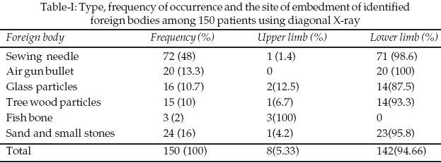
Sewing needle was the most common penetrating foreign body
and most affected limb was the right lower limb (Table-I) and the least
foreign body was fish bone, which was confined to females. Fifty one (34%)
cases were housewives and eighty six (57.3%) cases were young patients aged
less than 18 years old. Air gun bullet and tree wood particles were found in
twenty (13.3%) and fifteen (15%), respectively, were only seen in males
(Table-I). Glass particles were identified in five females and eleven males,
while sand and small stones were found in twenty and three cases of females
and one male respectively. In 147 cases (98%) the foreign body was removed in
the first surgical attempt, which normally lasted between 15 to 20 minutes. In
two failed cases (1.33%) were due to non-radio-opaque foreign bodies due to
wood and fishbone, which comprised 11% of total non-radio opaque objects. A
second surgical attempt was necessary for removal of these objects.
Fluoroscopy was employed for removal of a missed sewing needle (embedded in
the third metatarsal bone) case.
DISCUSSION
Retention of foreign bodies can lead to persistent
inflammation and infection. Early diagnosis and accurate localization of
foreign bodies is necessary for their successful removal.
7
Different diagnostic equipments are available for localizing foreign bodies.
Existence and localization of foreign bodies can be done by radiography,
ultrasonography, CT-Scan, MRI and intra-operative fluoroscopy.1
MRI, sonography and power doppler are the most sensitive, albeit expensive or
need trained staff, methods.2,3
In this three-year retrospective study, using two diagonally positioned plain
X-ray radiographies, 147 cases out of a total of 150 cases of radio-opaque and
non-radio-opaque foreign bodies were identified. Sonography had sensitivity of
92% for the overall
detection of foreign bodies,8
while power doppler imaging detected the foreign bodies in 100% of cases due
to its high sensitivity in detecting the high vascularity surrounding
chronically embedded foreign bodies.8
Although it is generally assumed that non-radio-opaque
subjects can not be detected by plain radiography, our study has demonstrated
that modifying this technique by taking two diagonally positioned x-ray
radiographs can ameliorate this shortfall. With careful medical history it can
successfully be used for detection of both radio-opaque and non radio-opaque
objects. The success rate of this technique was found to be 99.25% for
radio-opaque objects while for non radio-opaque objects this figure was 89%.
Furthermore, although CT and MRI can be used for the detection of wooden
foreign bodies in the extremities, they are more expensive than ultrasound and
are not easily available.
9,10
In conclusion, two diagonally positioned x-ray radiographs is highly sensitive
and accurate in the detection of radio-opaque and non radio-opaque objects in
the extremities. It is also useful in localizing and removal of foreign
bodies, thus minimizing dissection and operating time.
REFERENCES
1. Schmitt WG, Hubener HK. Demonstracton of non-metallic
foreign bodies by conventional X-Ray techniques and computer tomography.
Absorption coefficient of glass and wood. Rofo 1981;135(2):206-13.
2. Bray BW, Mahoney JL, Campbell JP. Sensitivity and
specificity of ultrasound in the diagnosis of foreign bodies in the hand. J
Hand Surg Am 1995;20:661.
3. Bodne D, Quinn SF, Cochram CF. Imaging foreign glass and
wooden bodies of the extremities with CT and MRI. J Comput Assist Tomogram
1988;12(4):608-11.
4. Cakr, Bars, Akan, Mithat, Serkan Y, Tayfun A.
Localization and removal of ferromagnetic foreign bodies by magnet. Annals
Plast Surg 2002;49:541.
5. Leung A, Patton A, Navoy J, Cummings RJ. Intraoperative
sonography-guided removal of radio lucent foreign bodies, J Ped Ortho
1998;18:259.
6. Lyno M, Brannam L, Johnson D, Blaivas M, Duggals.
Detection of soft tissue foreign bodies in the presence of soft tissue gas. J
Ultrasound Med 2004;23(5):677-81.
7. Flom LL, Ellis GL. Radio logic evaluation of foreign
bodies. Emerg Med Clin North Am 1992;10:163-77.
8. Davae KC, Sofka CM, DiCarlo E, Adler RS. Value of
power doppler imaging and the hypoechoic halo in the sonographic detection of
foreign bodies: Correlation with histopathologic findings. Ultrasound Med
2005;22:1309-13.
9. Bauer AR Jr, Yutani D. Computed tomographic localization
of wooden foreign bodies in children’s extremities. Arch Surg 1983;118:1084-6.
10. Bodne D, Quinn SF, Cochran CF. Imaging foreign glass and wooden bodies
of the extremities with CT 03-227-final 481 12/13/04, 10:36 AM and MR. J
Comput Assist Tomogr 1988;12:606-11.