|
 |
|
Published
by : PROFESSIONAL MEDICAL PUBLICATIONS |
|
ISSN 1681-715X |
|
|
|
- |
|
ORIGINAL
ARTICLE |
|
- |
|
Volume 25 |
October - December 2009
(Part-I) |
Number 5 |
|
|
|
Vitamin-C protect ethanol induced apoptotic
neurodegeneration in postnatal rat brain
Muhammad Imran Naseer1, Najeebullah2,
Ikramullah3, Hassan Zubair4,
Mukhtiar Hassan5, Byoung Chul Yang6, Myeong Ok Kim7
ABSTRACT
Objective: To evaluate ethanol effects to induced
activation of caspsae-3, and to observe the protective effects of Vitamin C (vit-C)
on ethanol-induced apoptotic neurodegeneration in rat cortical area of brain.
Methodology: Administration of a single dose of
ethanol in 7-d postnatal (P7) rats triggers activation of caspase-3 and
widespread apoptotic neuronal death. Western blot analysis, cells counting and
Nissl staining were used to elucidate possible protective effect of vit-C
against ethanol-induced apoptotic neurodegeneration in brain.
Results: The results showed that ethanol
significantly increased caspase-3 expression and neuronal apoptosis.
Furthermore, the co-treatment of vit-C along with ethanol showed significantly
decreased expression of caspase-3 as compare to control group.
Conclusion: Our findings indicate that vit-C can
prevent some of the deleterious effect of ethanol on developing rat brain when
given after ethanol exposure and can be used as an effective protective agent
for Fetal Alcohol Syndrome (FAS).
KEY WORDS:
Ethanol,
Neurodegeneration, Vitamin C, Cortex.
Pak J Med Sci October - December 2009
(Part-I) Vol. 25 No. 5 718-722
How to cite this article:
Naseer MI, Najeebullah, Ikramullah, Zubair H, Hassan M, Yang BC, et al.
Vitamin-C protect ethanol induced apoptotic neurodegeneration in postnatal rat
brain. Pak J Med Sci 2009;25(5):718-722.
1. Muhammad Imran Naseer,
2. Najeebullah,
* M. Imran Naseer & Najeebullah contributed
equaly to this work.
3. Ikramullah,
4. Hassan Zubair,
Sheffield Hallam University,
Sheffield, S1 1WB, United Kingdom
5. Mukhtiar Hassan
Hazara University Mansehra N.W.F.P. Pakistan
6. Byoung Chul Yang,
National Institute of Animal Science,
RDA Suwan, South Korea
7. Myeong Ok Kim,
1-3,7: Division of Life Science,
College of Natural Sciences and Applied Life Science
(Brain Korea 21), Gyeongsang National University,
Gazwa 351-301, Chinju 660-701, Republic of Korea.
Correspondence:
Prof. Myeong Ok Kim, Ph.D
E-mail: mokim@gsnu.ac.kr
* Received for Publication: August 4, 2009
* Accepted: September 15, 2009
INTRODUCTION
Ethanol is the most common human teratogen. Excessive
alcohol consumption during pregnancy can result in fetal alcohol syndrome (FAS).
It is characterized by abnormalities in the central nervous system (CNS).
These are reduced brain size (microencephaly), growth retardation, and facial
dysmorphology in the newborn children.
1
Ethanol may damage the developing brain by affecting neurogenesis, migration,
or survival of cells.2
Neurons are more susceptible to ethanol-induced apoptotic cell death during
synaptogenesis, also known as the brain growth spurt.3-4
Several studies have documented specific ethanol-induced
damage to the hippocampus. The hippocampal formation and number of neurons are
adversely affected by exposure to high doses of ethanol in neonatal rats.
5
Cell migration and differentiation are important processes for a developing
fetal central nervous system (CNS), and apoptosis is an indispensable
mechanism for these processes. Apoptosis discriminates the cell type,
population, and function in various regions.6-8
Previous studies have also shown that ethanol induces widespread apoptotic
neurodege-neration in the developing rat forebrain.3
Ascorbic acid concentration in brain is highly regulated.
The brain normally contains high concentrations of vit-C, which is actively
taken through the choroids plexus. However, its specific functions in the CNS
are only beginning to be elucidated. Vit-C acts as part of the intracellular
antioxidant network, and as such is an important neuroprotective constituent.
Recently, it has become clear that antioxidant nutrients, including vit-C, are
important for neurological function.
9-12
High intake of vitamin E and C has been found to be associated with lower risk
of Alzheimer’s disease.13
Therefore, the objective of this study was to assess the protective effect of
of vit-C against the ethanol mediated toxic effects relevant to neurological
damage.
Our results suggested that ethanol induced apoptotic
neurodegeneration, while the vit-C, may effectively protects against the
deleterious effects of ethanol-induced abnormalities, which may be used as a
therapeutic approach towards FAS-associated brain damage during early
developmental stages.
METHODOLOGY
Animals and drug treatment: P7 Sparague-Dawley rats of
15g (Gyeongsang National University, Neurobiology Laboratory, Chinju, South
Korea) were injected subcutaneously with 20% ethanol in saline solution
delivering 5g/kg body weight and 200mg/kg of vit-C and scarified after 4 h,
while during co treatment of vit-C plus ethanol, vit-C was injected after 30
min of ethanol treatment, whereas control group were treated with saline.
Cell Counting: Cells were counted one section out of
every six (240 ěm apart from each other) throughout the brain by using a Nikon
Eclipse E600 with a Nikon 40× objective. Two to four sections per brain for
the LDN (lateral dorsal nucleus) and six to ten sections per brain for the
cingulate cortex were counted in five to six animals of each group. Cells were
counted in both hemispheres. Contours of Cingulate CX (Bregma from 1.34 to
"0.82)
14
were traced using a personal computer, and the area was calculated with Stereo
Investigator software (MBF Bioscience, Williston, Vermont, United States).
Western blotting: Animals were killed at four hours
after ethanol and vit-C administration. Brains were dissected out and cortical
part was removed carefully and tissue was frozen in dry ice. For each
treatment group, three to four pups from 6 different litters were analyzed.
Caspase-3 analysis was done as previously described with some modification.
15
Western blots were analyzed by densitometry using the computer-based Sigma Gel
(SPSS Inc. Chicago, USA) system. Density values were expressed as mean ± SEM.
One-way ANOVA analysis followed by Tukey-Kramer multiple-comparisons test was
performed to determine the significance of differences between relevant
treatment groups. In every case, the acceptance level for statistical
significance was *P < 0.05.
Nissl staining: Nissl staining was done as previously
described with some modification.
16
Morphology of cells and presence of apoptotic and necrotic bodies was assessed
on microscope slides mounted 12µm thick brain sections. Cresyl violet,
recognize all structure particular nucleus and nucleic acids appear violet,
neurons faintly blue. Neuronal damage was then estimated as a rate of the
number of degenerated neurons to that of both surviving and degenerated in
three distinct areas of the cingulate cortex in coronal sections for each
animal.17
Data analysis and statistics: The object band from
Western blot were scanned and analyzed by densitometry using a computer based
on the Sigma Gel System (SPSS Inc., Chicago, IL). Density values were
expressed as mean ± SEM. One-way ANOVA analysis followed by Tukey–Kramer
multiple-comparisons test was performed to determine the significance of
differences between relevant treatment groups. The acceptance level for
statistical significance was *P < 0.05.
RESULTS
Vitamin C inhibits Ethanol-induced apoptotic
neurodegeneration: Activation of caspases resulted in nuclear,
plasma-membrane and mitochondrial changes. In the present study exposure of
ethanol significantly increased the expression of caspase-3. Increased
caspase-3 can disturb essential homeostatic processes and initiate an orderly
disassembly of cells including degradation of genomic DNA.
18
In order to determine that vit-C can inhibit ethanol-induced caspase-3
activation, a series of different experiments was performed. The doses of vit-C
200mg/kg were administered subcutaneously 30 minutes after ethanol treatment,
Western blot results showed that the animals injected with ethanol alone
showed a 10-fold to 20-fold increase of caspase-3 compared with a saline
control, while the co treatment of vit-C with ethanol significantly decreased
the expression of caspase-3 as compare to ethanol treated group (Figure 1).
The high concentrations of ethanol were used in the experiment to evaluate the
effect of ethanol on developing brain (Fig- 1).

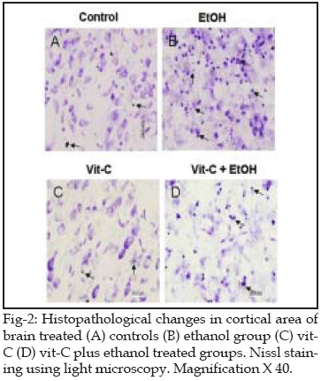
Histological findings: To determine whether inhibition
of caspase-3 activation by vit-c is sufficient to prevent ethanol-induced cell
death, brains were histologically analyzed for evidence of neurodegeneration.
The Nissl staining results showed that ethanol significantly increased
neuronal death, while the co treatment of vit-C with ethanol significantly
inhibited the neuronal death compared to ethanol treated group (Fig- 2).
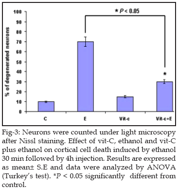
Which suggest that vit-C an antioxidant, may effectively
protects against the deleterious effects of ethanol-induced abnormalities by
decreasing neuronal death in cortical rat brain, furthermore the cell counting
under light microscope after Nissl stain also showed the same increased
neurodegeneration upon ethanol treated group and decreased significantly when
treated with vit-c as compare to alone ethanol treated group (Fig- 3).
DISCUSSION
Brain damage due to ethanol exposure is a cardinal feature
in (FAS) in alcoholism. Enhanced neurodegeneration
3
in combination with oxidative stress outcomes probably lead to the
neurodevelopmental deficiencies that are encountered in FAS. It has already
been established that ethanol decreases the population of neurons in the brain
and causes malformation including the loss of brain mass.19-21
Present study indicate the apoptotic neurodegeneration induced by ethanol and
a protective effect of antioxidants vit-C during early developmental stage.
Cell proliferation and differentiation are two critical
processes in a developing fetal brain. Apoptosis Bcl-2 family proteins, plays
decisive role.
22-24
Moreover, the normal population of neurons is controlled by a balance of
apoptosis and stem cell activities. In the present study, the activation of
caspase-3 in response to ethanol exposure was investigated using Western blot
analysis. It revealed increase expression of caspase-3 proteins. Furthermore,
we have also demonstrated that the co administration of vit-C with ethanol
treatment in early postnatal development prevents alcohol-induced apoptotic
neurodegeneration. Administration of vit-C protected against this
ethanol-induced apoptosis, suggests a link between neuronal loss during brain
development and behavioral disturbances observed in the adult rat.
Ethanol exposure during prenatal development causes wide
range of structural and functional brain abnormalities resulting in the
condition of fetal alcohol effect (FAE) and alcohol related neurodevelopmental
disorders (ARNDS) including cytoarchitectual abnormalities.
25-26
In this study, we showed that the
antioxidant vit-C can effectively reduce the severity of ethanol-induced brain
injury and growth retardation during early brain development. The antioxidant
vit-C can effectively reduce the severity of ethanol-induced brain injury and
growth retardation. It is conceivable that vit-C can exert its neuroprotective
role as a potent scavenger of oxygen free radicals. Second, it restores the
expression of important neural markers including NCAM and Pax6. Third, it may
also interact with ethanol extracellularly and hence alleviate the overall
teratogenic effect of ethanol.27
The mechanisms underlying the vit-c neuroprotective effects are not fully
understood, however, our results showed that co treatment of vit-C plus
ethanol are in agreement with neuroprotective actions of vit-c reported in
previous studies by pilocarpine.28
In summary, this study showed that ethanol induced
apoptosis neurodegeneration and the co treatment of vit-C with ethanol
decreased ethanol-induced apoptotic neurodegeneration in developing brain.
Although additional studies are necessary, we suggest, that vit-C a readily
available and safe agent, could be used for the treatment and prevention of
the FAS.
ACKNOWLEDGEMENT
This work was supported by KOSEF, grant funded by the
Korean government (2009-0058805) Brain Korea 21 and a grant No.
20080401034062, from BioGreen 21 program.
REFERENCES
1. Stratton K, Howe C, Battaglia F. Fetal Ethanol Syndrome:
Diagnosis, Epidemiology, Prevention, and Treatment. Washington, DC. National
Academy Press, (Eds.), 1996.
2. Miller, M.W. Mechanisms of ethanol induced neuronal
death during development: from the molecule to behavior. Alcohol Clin Exp Res
1996;20:128–132.
3. Ikonomidou CP, Bittigau MJ, Ishimaru DF, Wozniak C, Koch
K, Genz MT. et al Ethanol-induced apoptotic neurodegeneration and fetal
alcohol syndrome. Science 2000;287:1056–1060.
4. Olney JW, Ishimaru MJ, Bittigau P, Ikonomidou C.
Ethanol-induced apoptotic neurodegeneration in the developing brain. Apoptosis
2000;5:515–521
5. Miki T, Harris SJ, Wilce P, Takeuchi Y, Bedi KS. Neurons
in the hilus region of the rat hippocampus are depleted in number by exposure
to alcohol during early postnatal life. Hippocampus 2000;10:284–295.
6. Kuan C. Mechanisms of programmed cell death in the
developing brain. Trends Neurosci 2000;23:291-297.
7. Kuida K, Zheng TS, Na S, Kuan C, Yang D, Karasuyama H.
et al Decreased apoptosis in the brain and premature lethality in CPP32-
deficient mice. Nature 1996;384:368-372.
8. Yoshida H, Kong YY, Yoshida R, Elia AJ, Hakem A, Hakem
R, et al. Apaf1 is required for mitochondrial pathways of apoptosis and brain
development. Cell 1998;94:739-750.
9. Daskalopoulos R, Korcok, J, Tao L, Wilson JX.
Accumulation of intracellular ascorbate from dehydroascorbic acid by
astrocytes is decreased after oxidative stress and restored by propofol. Glia
2002;39:124–32.
10. Rice ME. Ascorbate regulation and its neuroprotective
role in the brain. Trends Neurosci 2000;23:209–16.
11. Siushansian R, Wilson JX. Ascorbate transport and
intracellular concentration in cerebral astrocytes. J Neurochem 1995;65:41–9.
12. Wilson JX, Peters CE, Sitar SM, Daoust P, Gelb AW.
Glutamate stimulates ascorbate transport by astrocytes. Brain Res
2000;858:61–6.
13. Engelhart MJ, Geerlings MI, Ruitenberg A, van Swieten
JC, Hofman A, Witteman JC, et al. Dietary intake of antioxidants and risk of
Alzheimer disease. JAMA 2002;287:3223–9.
14. Paxinos G, Franklin KBJ. The mouse brain in stereotaxic
coordinates. San Diego: Academic Press; 186 pp 1997.
15. Naseer MI, Li SP, Kim MO. Maternal epileptic seizure
induced by Pentylenetetrazol: Apoptotic neurodegeneration and decreased GABAB1
receptor expression in prenatal rat brain. Molecular brain 2009;2(1):20.
16. Majid AS, Marzieh P, Shahriar SK, Pari KT.
Neuroprotective effects of Aqueous Date Fruit Extract on focal cerebral
ischemia in rats. Pak J Med Sci 2008;24(5):661-65.
17. Sakurai-Yamashita Y, Kinugawa H, Niwa M.
Neuroprotective effect of pentosan polysulphate on ischemia-related neuronal
death of hippocampus. Neurosci Lett 2006;409:30-4.
18. Zhu C, Wang X, Hagberg H, Blomgren K. Correlation
between caspase-3 activation and three different markers of DNA damage in
neonatal–cerebral hypoxia–ischemia. J Neurochem 2000;75:819–829.
19. Jones KL, Smith DW, Ulleland CN, Streissguth AP.
Pattern of malformation in offspring of chronic alcoholic mothers. Lancet
1973;1:1267-1271.
20. Miki T, Harris SJ, Wilce PA, Takeuchi Y, Bedi KS.
Effects of alcohol exposure during early life on neuron numbers in the rat
hippocampus, I. Hilus neurons and granule cells. Hippocampus 2001;13:388-398.
21. Sakata-Haga H, Sawada K, Hisano S, Fukui Y.
Administration schedule for an ethanol-containing diet in pregnancy affects
types of offspring brain malformations. Acta Neuropathol 2002;104:305-312.
22. Bibel M, Barde YA. Neurotrophins: Key regulators of
cell fate and cell shape in the vertebrate nervous system. Genes Dev
2000;14:2919-2937.
23. Montoliu C, Valles S, Renau-Piqueras J, Guerri C.
Ethanol-induced oxygen radical formation and lipid peroxidation in rat brain:
Effect of chronic alcohol consumption. J. Neurochem 1994;63:1855-1862.
24. Dunty JWC, Chen SY, Zucker RM, Dehart DB, Sulik KK.
Selective vulnerability of embryonic cell populations to ethanol-induced
apoptosis: Implications for alcohol-related birth defects and
neurodevelopmental disorder. Alcohol Clin Exp Res 2001;25:1523-1535.
25. Clarren, SK, Smith DW. The fetal alcohol syndrome. New
Eng J Med 1978;298:1063-1067.
26. Lindsley TA, Kerlin AM, Rising LJ. Time-lapse analysis
of ethanol’s effects on axon growth in vitro. Dev Brain Res 2003;30:191-199.
27. Penga Y, Kwok KHH, Yang PH, Samuel SMN, Liu J, Wong OG,
et al. Ascorbic acid inhibits ROS production, NF-kB activation and prevents
ethanol-induced growth retardation and microencephaly. Neuropharmacology
2005;48:426–434.
28. Xavier SM, Barbosa CO, Barros DO, Silva RF, Oliveira AA, Freitas RM.
Vitamin C antioxidante effects in hippocampus of adult Wistar rats after
seizures and status epilepticus induced by pilocarpine. Neurosci Lett
2007;8:76–79.
HOME
| SEARCH
| CURRENT
ISSUE | PAST
ISSUES
Professional
Medical Publications
Room No. 522, 5th Floor, Panorama Centre
Building No. 2, P.O. Box 8766, Saddar, Karachi - Pakistan.
Phones : 5688791, 5689285 Fax : 5689860
pjms@pjms.com.pk