|
|
||||
|
Published by : PROFESSIONAL MEDICAL PUBLICATIONS |
||||
|
ISSN 1681-715X |
||||
|
||||
|
- |
||||
|
ORIGINAL ARTICLE- |
||||
|
- |
||||
|
Volume 23 |
April - June 2007 (Part-I) |
Number 2 |
||
|
|
||||
|
|
||||
|
|
||||
|
Published by : PROFESSIONAL MEDICAL PUBLICATIONS |
||||
|
ISSN 1681-715X |
||||
|
||||
|
- |
||||
|
ORIGINAL ARTICLE- |
||||
|
- |
||||
|
Volume 23 |
April - June 2007 (Part-I) |
Number 2 |
||
|
|
||||
|
|
||||
Urogenital anomalies associated with
anorectal malformations
Fakhrossadat Mortazavi1, Saeid Aslanabadi2, Seyyedeh Tayyebeh Mahnama3ABSTRACT
Objective: Urogenital anomalies are frequently associated with anorectal malformations which are a common source of significant morbidity. The aim of this study was to evaluate the incidence and nature of associated urogenital anomalies in patients with Anorectal Malformations (ARM).
Methodology: Documents of 104 patients with ARM were studied from 2003 to 2005. All patients underwent sonography of urinary tract and lumbosacral radiography. Other imaging studies were done in selected cases and voiding cystourethrography (VCUG) performed in 62 patients.
Results: Urologic malformations were found in 44 patients (42.3%) without sexual preponderance (p>0.05). Genital anomalies were detected in 16 cases (15.5%) with a significantly high frequency in males (p<0.05). The incidence of associated urogenital anomalies was significantly higher in “high” form of anomaly than those with “low” form (p<0.05). Vesicoureteral reflux (VUR), hydronephrosis, and renal agenesis were the most common urologic anomalies respectively. Cryptorchidism and hypospadiasis were the most frequent genital anomalies. Sacrospinal anomalies were detected in 22% of patients.
Conclusion: The high incidence of associated urogenital anomalies necessitates a careful investigation of all patients with ARM. VCUG is essential even with normal sonographic findings.
KEY WORDS: Anorectal malformations, Urinary tract, Genital, Urogenital, Association.
Pak J Med Sci April 2007 Vol. 23 No. 2 172-175
1. Dr. Fakhrossadat Mortazavi,
Department of Pediatric Nephrology,
2. Dr. Saeid Aslanabadi,
Department of Pediatric Surgery,
3. Dr. Seyyedeh Tayyebeh Mahnama,
Department of Pediatrics,
1-2: Assistant Professor,
Tabriz University of Medical Science,
Faculty of Medicine,
Children’s Hospital of Tabriz,
Tabriz – Iran.
Correspondence:
Dr. Fakhrossadat Mortazavi,
Children’s Hospital of Tabriz,
Sheshgelan Street,
Tabriz – Iran.
E-Mail: fmortazavi@tbzmed.ac.ir
* Received for Publication: July 25, 2006
* Accepted: October 4, 2006
INTRODUCTION
Anorectal malformations (ARM) occurs in one of every 4000-5000 newborns and incidence is slightly higher in male than female.1-3 Several classification systems of this defect have been proposed in the surgical literature. ARM may be classified as either “high” or “low” anomalies based on their relationship to the levator muscle complex.2-4 About 50-60% of infants have associated malformations including urogenital, cardiovascular, sacrospinal and gastrointestinal anomalies.1-5 Occasionally patients present with a constellation of anomalies as a part of the “VACTERL” association (Vertebral, Anorectal, Cardiac, Tracheoesophageal, Renal, Radial and Limb anomalies).4-6,7 Associated anomalies are more common in high than low level deformities.5 The true incidence of genitourinary anomalies found in patients with ARM may only be detected by complete urologic evaluation. Urologic malformations may cause renal damage and may lead to chromic renal failure if not detected in time. Early diagnosis of urogenital malformations in neonatal period is essential in preventing future complications. The objective of this paper is to review the incidence and types of associated urogenital anomalies with ARM in our institution and compare the results with other similar studies.
PATIENTS AND METHODS
In this cross sectional study, we reviewed the records of 104 neonates with ARM who were admitted in Children’s Hospital of Tabriz, between April 2003 and March 2005. The variables included: sex, level of imperforate anus (High or low), findings in physical examination, and the results of imaging studies. The level of anomaly was detected by radiography and surgeon’s report on operation. All patients underwent ultrasound evaluation of the urinary tract, AP and lateral X-ray of lumbosacral spine. Renal isotope scan and intravenous urography were done in selected cases. Patients with abnormal sonography underwent VCUG in neonatal period and those with normal sonography had VCUG in outpatient follow up. At the time of review the results of VCUG were available in sixty two patients. Urinary tract anomalies were defined as any renal, ureteral or bladder malformation, excluding rectovesical and rectourethral fistula. Genital anomalies were diagnosed by physical examination followed by imaging studies when needed. Grading of reflux was done according to international classification. The data were analyzed by chi-square and fisher’s exact test and differences considered statistically significant for P values < 0.05.
RESULTS
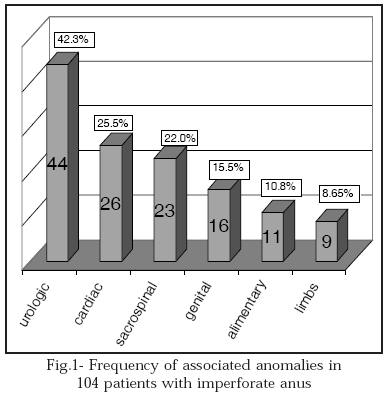
Of the 104 patients with imperforate anus 58(55.8%) were male (38 with high and 20 with low type deformity) and 46 (44.2%) were female (24 with high and 22 with low type deformity). There was no significant difference between boys and girls for level of imperforate anus (P=0.168). At least one associated anomaly was detected in 71(68.2%) patients (50 of them with high and 21 with low type of deformity) with a preponderance in “high” level deformities than “low” ones (P= 0.002). Urologic, cardiac and sacrospinal anomalies were the most frequent associated anomalies respectively (Fig-1).
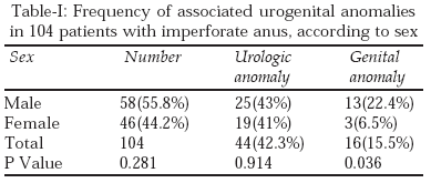
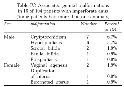
Forty-four of 104 (42.3%) patients had at least one urologic malformation with no sexual preponderance and 16 (15.3%) patients had genital malformation with significantly high incidence in males. Table-I.
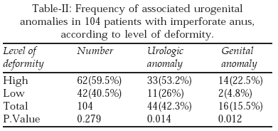
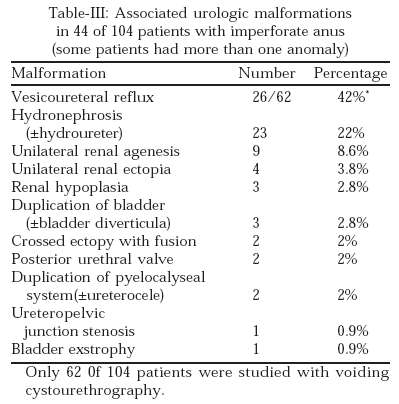
The Incidence of associated urogenital anomalies was higher significantly more common in patients with “high” type ARM (Table-V). Vesicoureteral Reflux (VUR), hydronephrosis and unilateral renal agenesis were the most common urologic anomalies respectively (Table-III).
Cryptorchidism and hypospadiasis were the most common genital anomalies (Table-IV). VUR was found in 26 of 62 (42%) patients investigated by VCUG. VUR was bilateral in eight and unilateral in 18 cases. So there were a total of 34 refluxing renal units. Grade I was seen in five, grade II in nine, grade III in eleven, grade IV in five and grade V in 4 kidneys. In 11 of 26 patients with VUR (42.3%), sonography of urinary tract was normal. In 7 of 15 patients with unilateral renal ectopia, hypoplasia and agenesis, VUR was found in contra lateral kidney (7/15=46.6%). Hydronephrosis was detected in 23 (22%) patients, accompanied with hydroureter in fifteen cases.
The most common cause of hydronephrosis was VUR which was present in 11 of 23 patients. Sacrospinal anomalies were detected in 23 (22%) patients (19 with “high” and four with “low” level lesions), among these hemivertebra was the most frequent anomaly. VACTERL association was present in two patients, and VATER and VACTER each in one patient (4/104 =3.8%).
DISCUSSION
Urinary tract abnormalities are the most common associated anomaly in patients with ARM and have been reported in 26-52% of several large series,7-10 Its incidence is higher in infants with a “high” versus a “low” anomaly and boys are more prone than girls to have an urologic anomalies.7,8 About 42.3% of our cases had urinary tract anomalies with a preponderance in high level deformities, but we didn’t find statistically significant difference in incidence of urologic anomalies, between males and females. VUR and renal agenesis are the most common associated urinary tract anomaly with imperforate anus.11,12 Metts and boemers found VUR in 32% and 27% of their cases respectively,8,11 Misra et al reported that 37.5% of patients with low deformity had VUR,13 but Rattan and Srivastava reported the incidence of VUR only in 1.7% and 5.4% of their patients.14-15 This wide variation in incidence of VUR is related to the different methods of studies. In some studies VCUG was performed only when sonographic findings were abnormal. In this study, although VCUG was performed only in 62 of 104 patients, but twenty six had VUR showing higher incidence than other anomalies. Hydronephrosis and renal agenesis are the most common anomalies of the upper urinary tract in this study. Considering that hydronephrosis is secondary to other anomalies such as VUR and bladder dysfunction, renal agenesis may be considered as the most common primary anomaly of upper urinary tract, similar to literature.
Neurovesical dysfunction (NVD) is a frequent finding in children with anorectal malformatins. Emir et al. reported its incidence 45.4%.16 Boemers found NVD in 24% of cases.11 Sheldon et al reported NVD in 70% of patients with imperforate anus, who underwent genitourinary procedures.17 NVD commonly is associated with sacrospinal deformities but some authors recommend evaluation of all patients with magnetic resonance imaging (MRI) because spinal cord anomalies may occur without obvious sacrospinal anomalies.18 Urodynamic studies (UDS) are reserved for those children with either a deformity of the spine or a spinal cord defect or any signs of NVD on a VCUG and sonography.7,18 In this study 22% of patients had bony sacrospinal deformity, but they were not investigated for NVD. In future studies we have to consider MRI and UDS for evaluation of NVD in our patients.
In this study the incidence of genital anomalies was 22.4% in boys and 6.5% in girls with an overall incidence of 15.5%, similar to the 16.4% and 16.5% reported by Metts and Mclorie respectively.8 In our patients cryptorchidism and hypospadias were the most common genital anomalies, as in cases of Metts. In some studies hypospadiasis has been reported as most common genital anomaly.15 A lower incidence of genital anomalies in girls could be attributed to an inadvertent missing of internal genitalia abnormalities in the presence of normal looking external genitalia.
In conclusion all patients with imperforate anus should be thoroughly investigated for urogenital and spinal anomalies. VCUG is mandatory even in those with normal sonography. Prophylaxis for urinary infection should be initiated until VCUG is performed, because the incidence of VUR is really high. Appropriate urologic care is essential to prevent renal damage.REFERENCES
1. Pena A. Imperforate anus and cloacal malformations. In: Ashcraft KW, Murphy S. Pediatric surgery. 3rd ed. Philadelphia; Saunders 2002;p.473-7.
2. Kiely EM, Pena A. Anorectal malformation. In: o‘neil JA, Rowe MI, Grosfeld JL, Fonkalsrud EW, Coran AG. Pediatric surgery. 5thedition. St. Louis; Mosby 1998;p.1425-46.
3. O‘Neil JA, Grosfeld JL, Fonkalsrud EW, Coran AG, Caldamone AC. Anorectal disorders and imperforate anus. In: Principles of pediatric surgery. 2ndedition. New York; Mosby 2004;p.596.
4. Bullard KM, Rothenberger DA. Colon, Rectum and Anus. In: Brunicardi FC, Andersen DK, Billiar TR, Hunter JG. Schwartz’s principle of surgery. 8th edition. New York; McGraw-Hill 2005;p.1497-9.
5. Endo M, Hayashi A, Ishihara M, Maie M, Nagasaki A, Nishi T , et al. Analysis of 1992 Patients with anorectal malformations over the past two decades in Japan. J Pediatr Surg 1999;vol 34(3):435-41.
6. Miller OF, Kolon TF. Prenatal diagnosis of VACTERL association. J Urol 2001;166:2389-91.
7. Bauer SB, Koff SA, Jayanthi VR. Voiding dysfunction in children: neurogenic and non-neurogenic. In: Walsh PC, Retic AB, Vaughan ED, Wein AJ. Campbells Urology. 8th edition. Philadelphia; Saunders 2002;p.2250-4.
8. Metts JC, Kotkin L, kasper S, Shyr YU, Adams MC, Brock JW. Genital malformations and coexistent urinary tract or spinal anomalies in patients with imperforate anus. J Urol 1997;158:1298-300.
9. Marlinez–Frias ML, Bermejo E, Rodriguez–pinilla E. Anal atresia, vertebral, genital and urinary tract anomalies: A primary polytopic developmental field defect identified through an epidemiological analysis of associations. Am J Med Genet 2000;13:95(2):169-73.
10. Jun W, Chengren S, Shiyao Y, Yan W, Changhui X. A rare association of rectal and genitourinary duplication and anorectal malformation. Chin Med J 2003;116(12):1955-7.
11. Boemers TML, Jong TPVM, Van Gool JD, Bax KMA. Urologic problems in anorectal malformations part 2: Functional urologic sequelae. J Pediatr Surg 1996; 31(5):634-7.
12. Banever GT, Moriarty KP. Posterior urethral valve in a newborn with imperforate anus: clinical presentation and management. J Pediatr Surg 2005;40:1332-4.
13. Misra D, Mushtaq I, Dpake DP, Kiely EM, Spitz L. Associated urologic anomalies in low imperforate anus are capable of causing significant morbidity. J Urol 1996;48:281-3.
14. Ratan SK, Rattan KN, Pandey RM, Mittal A, Magu S, Sodhi PK. Associated congenital anomalies in patients with anorectal malformations-A need for developing a uniform practical approach. J Pediatr Surg 2004;vol 39(11):1706-11.
15. Srivastava V, Ray AK, Patra R, Saha BK, Samanta N, Saha K. Urogenital anomalies associated with anorectal malformation. J Indian Assoc Pediatr Surg 2005;10:44-7.
16. Emir H, Soylet Y. Neurovesical dysfunction in patients with anorectal malformations. Eur J Pediatr Surg 1999;8(2):95-7.
17. Sheldon CA, Gilbert A, Lewis AG, Aiken J, Ziegler MM. Surgical implications of genitourinary tract anomalies in patients with imperforate anus. J Urol 1994;152(1):196-9.
18. Mosiello G, Capitanucci ML, Gatti C, Adorisio O, Lucchetti MC, Silveri M. How to investigate neurovesical dysfunction in children with anorectal malformations. J Urol 2003;170 (4):1610-3.
HOME | SEARCH | CURRENT ISSUE | PAST ISSUES
Professional
Medical Publications
Room No. 522, 5th Floor, Panorama Centre
Building No. 2, P.O. Box 8766, Saddar, Karachi - Pakistan.
Phones : 5688791, 5689285 Fax : 5689860
pjms@pjms.com.pk