|
|
||||
|
Published by : PROFESSIONAL MEDICAL PUBLICATIONS |
||||
|
ISSN 1681-715X |
||||
|
||||
|
- |
||||
|
ORIGINAL ARTICLE |
||||
|
- |
||||
|
Volume 24 |
January - March 2008 |
Number 1 |
||
|
|
||||
|
|
||||
|
|
||||
|
Published by : PROFESSIONAL MEDICAL PUBLICATIONS |
||||
|
ISSN 1681-715X |
||||
|
||||
|
- |
||||
|
ORIGINAL ARTICLE |
||||
|
- |
||||
|
Volume 24 |
January - March 2008 |
Number 1 |
||
|
|
||||
|
|
||||
Hysteroscopic View of
Endometrial HyperplasiaMojgan Barati1, Sara Masihi2, Farideh Moramezi3
ABSTRACT
Objective: To determine the hysteroscopic appearance of endometrial hyperplasia in women with subsequently confirmed diagnosis of endometrial hyperplasia.
Methodology: This study was done in Aria Hospital in Ahwaz,Iran fromJanuary 21, 2003 to May 24th 2005. Fifty women underwent hysteroscopy with eye direct biopsy of the endometrium. Cause of hysteroscopy was Abnormal Uterine Bleeding (AUB) in 93.5% of cases. Specimens were sent for histology assessment. From these patients five cases had pathologic diagnosis of endometrial hyperplasia. Hysteroscopic features of these five cases were reviewed.
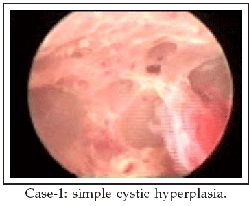
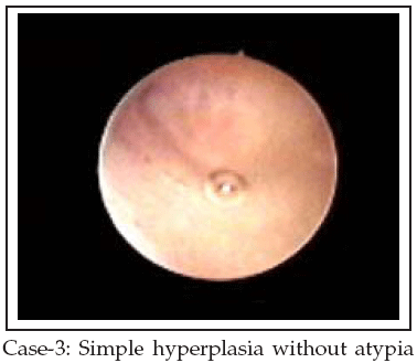
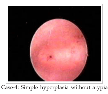
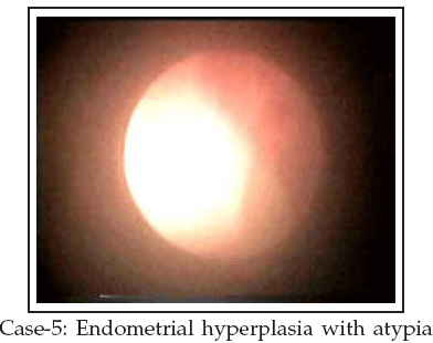
Results: In case one which was simple cystic hyperplasia, there was obvious cystic bizarre view. In case two, three and four there were minimal hysteroscopic abnormal view. In case 5 which was endometrial hyperplasia with atypia, there were obvious white endometrial elevations in the endometrial lining.
Conclusions: Endometrial hyperplasia may produce obvious space occupying lesions in which diagnosis is easy with hysteroscopy, but it may be not very obvious especially in early stages of the disease. In all these 5 cases there were white areas with markedly reduced or absent vascularity.
KEY WORDS: Hysteroscopy, Endometrial hyperplasia, White lesion.
Pak J Med Sci January - March 2008 Vol. 24 No. 1 65-68
1. Dr. Mojgan Barati MD,
2. Dr. Sara Masihi MD,
3. Dr. Farideh Moramezi MD,
1-3: Ahwaz University of Medical Science,
Ob&Gyn ward/ Emam Khomaini Hospital/ 24 metry
Ahwaz – Iran.
Correspondence
Dr. Mojgan Barati,
E-Mail: brati_m@yahoo.com
* Received for Publication: April 30, 2007
* Accepted for Publication: January 8, 2008
INTRODUCTION
Endometrial hyperplasia may be a precursor to the most common female genital malignancy, endometrial carcinoma. Unopposed estrogens from anovulatory cycles and exogenous use in postmenopausal women have been shown to increase the likelihood of endometrial hyperplasia and endometrial carcinoma.
1-4 Progression of endometrial hyperplasia to more aggressive pathology is time related. Simple hyperplasia often regresses if the source of exogenous estrogen is removed. However, atypical hyperplasia often progresses to adenocarcinoma unless medical intervention occurs.5 Less than 2% of hyperplasias without atypia progress to carcinoma, and the mean duration of progression to carcinoma take almost 10 years. Atypical hyperplasia progresses to carcinoma in 23% of cases over a mean duration of four years.6 Postmenopausal patients with endometrial hyperplasia invariably present with vaginal bleeding. Although carcinoma must be considered in this age group, endometrial atrophy represents the most common cause of postmenopausal bleeding. In a study of 226 women with postmenopausal bleeding, 7% were found to have carcinoma, 56% were noted to have atrophy, and 15% were diagnosed with some form of hyperplasia.7 Hyperplasia and carcinoma may present with heavy vaginal bleeding, whereas patients with atrophy usually present with light spotting.8 Meta analyses restricted to postmenopausal women with abnormal bleeding show that a positive test result following hysteroscopy is more useful for predicting endometrial cancer or hyperplasia disease than TVS.9 In contrast, a negative test result following Trans Vaginal Ultrasound (TVS) in postmenopausal women (4 or 5 mm cut-off to define abnormality) is highly accurate in excluding serious endometrial disease and more useful than hysteroscopy in this context.10,11 Applying the accuracy estimates from all three TVS reviews10-12 assuming a 5% pretest probability of cancer and endometrial thickness cut-offs of 4 or 5mm, the positive probability of cancer following a negative TVS is between 0.4 and 0.8%. The corresponding probability of cancer is 80% following a positive hysteroscopy.9 Endometrial thickness measurement using ultrasound is of minimal use in premenopausal women because specific cut-off levels or morphological features do not accurately define the presence or absence of endometrial hyperplasia or cancer.13 Outpatient endometrial biopsy has high accuracy in diagnosing endometrial cancer and hyperplasia and should be employed when serious endometrial disease is suspected in both pre and postmenopausal women.9 For many years dilatation and curettage (D&C) under general anesthesia was considered the gold standard for determining the cause of abnormal uterine bleeding.14 Less-invasive outpatients’ methods, such as Vabra and Pipelle, have similar or worse diagnostic accuracy, due to blind endometrial sampling.15 At the beginning of the 1990s, transvaginal sonography greatly improved the accuracy of evaluations of endometrial morphology, whereas in the last 10 years hysteroscopy has become, in some hospitals, the gold standard procedure for evaluating the uterine cavity, particularly if performed in an office setting and if associated with eye-guided biopsies.15-22 Hysteroscopy without endometrial biopsy is unreliable in differentiating between pre-malignant and malignant disease in the uterine cavity,23 although if the cavity is clearly atrophic it may be possible to omit endometrial sampling.24 Endometrial cancer may be found in symptomatic and asymptomatic women with an essentially atrophic or focally hyperplastic endometrium,25,26 which cannot be detected by ultrasound.In this study, we tried to describe hysteroscopic features of endometrial hyperplasia.
Patients AND METHODS
This study was done in Aria Hospital in Ahvaz, Iran from January 21, 2003 to May 24, 2005. In all fifty women underwent hysteroscopy. Cause of hysteroscopy was AUB, pain, asymptomatic and miscellaneous. Hysteroscopy directed biopsies were taken and specimens were sent for pathologic assessment. From these patients five cases had pathologic diagnosis of endometrial hyperplasia. Hysteroscopic features of these five cases were reviewed repeatedly.
RESULTS
In case one which was simple cystic hyperplasia, there was obvious cystic bizarre view. Panoramic view of endometrial cavity was distorted. Back to back areas of cystic figures in endometrium were seen. These cystic figures have white color.
In case two and three there were white suspicious lesions especially near cornea. Panoramic views were not distorted. Pathology report was hyperplasia without atypia in these two cases.
In case four, endometrium had atrophic appearance, panoramic view was not distorted. There was no suspicious area but now that we know the pathology report, near the cornea in fundal area there is diffuse white area. Pathology report was hyperplasia without atypia.
In case five which was endometrial hyperplasia with atypia, there were obvious white endometrial elevations. These elevations were friable and shiny white.
DISCUSSION
Endometrial hyperplasia may produce obvious intracavitary lesions which can be seen on hysteroscopy. These obvious intracavitary lesions are white, friable and little or no vessels are seen on them. These lesions may distort panoramic view of endometrial cavity. Also endometrial hyperplasia may exist in spite of little or no obvious endometrial lesions in hysteroscopy. It is doubtless that under direct eye vision, diagnosis of obvious space occupying lesions and taking biopsy is easy as in case one and five. In case two and three there were suspicious lesions near cornea. It seems that hyperplasia appears first in cornea. But one of the pit falls of hysteroscopy is pathologic diagnosis of hyperplasia in patients which hysteroscopic views were not having obvious lesions as in case four. So it is better to take biopsy in spite of normal hysteroscopic view.
In Rivierenland Hospital of Netherlands, de Wit AC, Vleugels MP, de Kruif JH performed 1045 diagnostic hysteroscopic procedures throughout six consecutive years, focusing on its value in diagnosing endometrial hyperplasia and carcinoma. They concluded that diagnostic hysteroscopy is a valuable diagnostic tool in diagnosing structural intra-cavital pathology, for the outpatient clinic. The value in diagnosing hyperplasia or endometrial carcinoma is limited and even after guided biopsy a malignancy cannot be ruled out.
27 G.Benagiano from Switzerland and L.Mencaglia from Italy have reported that there is no specific appearance for each histological type of endometrial hyperplasia. The hysteroscopic appearance of low risk endometrial hyperplasia (EH) includes an increase in the thickness of the endometrium, its dyshomo- geneous regeneration, increased vascularization and the presence of ciliated images, cystic dilatation, increased bleeding, polypoid formation, necrotic zones and the concentration and irregular arrangement of the glandular openings. In its initial stages, endometrial cancer shows a papillary appearance with irregular polylobate excrescences which are friable and partly necrotic or haemorrhagic. Vascularization is irregular and anarchic. Often there is a clear dividing line between cancerous and normal endometrium. Neoplastic lesions can be focal and localized at the tubal cornea.28 Overall, endometrial hyperplasia may produce obvious space occupying lesions in which diagnosis is easy with hysteroscopy, but it may not be very obvious especially in early stages of the disease. For determining hysteroscopic characteristics of these hidden hyperplasias more studies are necessary. However the final diagnosis of exact pathology depend on histopathology examination.REFERENCES
1. Antunes CMF, Stolley PD, Rosenshein NB. Endometrial cancer and estrogen use. Report of a large case-control study. N Engl J Med 1979;300:9-13.
2. Herrinton LJ, Weiss NS. Postmenopausal unopposed estrogen characteristics of use in relation to the risk of endometrial carcinoma. Ann Epidemiol 1993;3:308-18.
3. Jick H, Watkins RN, Hunter J. Replacement estrogens and endometrial cancer. N Engl J Med 1979;300:218-22.
4. Shapiro S, Kaufan DW, Slone E. Recent and past use of conjugated estrogens in relation to adenocarcinoma of the endometrium. N Engl J Med 1980;303:485-89.
5. Terakawa N, Kigawa J, Taketani Y. The behavior of endometrial hyperplasia: a prospective study. Endometrial Hyperplasia Study Group. J Obstet Gynaecol Res 1997;23:223-30.
6. Kurman RJ, Kaminski PF, Norris HJ. The behavior of endometrial hyperplasia. A long term study of untreated hy- perplasia in 170 patients. Cancer (Phila) 1985;56:403-12.
7. Lidor A, Ismajovich B, Condino E. Histopathologic findings in 226 women with postmenopausal uterine bleeding.Acta Obstet Gynecol Scand 1986;65:41-3.
8. Ronnett BM, Kurman RJ. Precursor lesions of endometrial carcinoma. In: Kurman RJ, ed. Blaustein’s Pathology of the Female Genital Tract, 5th ed. New York: Springer-Verlag 2002;467-500.
9. Clark TJ, Voit D, Gupta JK. Accuracy of hysteroscopy in the diagnosis of endometrial cancer and hyperplasia: A systematic quantitative review. JAMA 2002;288:1610-21.
10. Smith-Bindman R, Kerlikowske K, Feldstein VA. Endo- vaginal ultrasound to exclude endometrial cancer and other endometrial abnormalities. JAMA 1998;280:1510-17.
11. Gupta JK, Chien PF, Voit D. Ultrasonographic endometrial thickness for diagnosing endometrial pathology in women with postmenopausal bleeding: A meta-analysis. Acta Obstet Gynecol Scand 2002;81:799-816.
12. Tabor A, Watt HC, Wald NJ. Endometrial thickness as a test for endometrial cancer in women with postmenopausal vaginal bleeding. Obstet Gynecol 2002;99:663-70.
13. Farquhar C, Ekeroma A, Furness S, Arroll B. A systematic review of transvaginal ultrasonography, sonohysterography and hysteroscopy for the investigation of abnormal uterine bleeding in premenopausal women. Acta Obstet Gynecol Scand 2003;82:493-504.
14. Grimes DA. Diagnostic dilatation and curettage: A reappraisal. Am J Obstet Gynecol 1982; 142:1–6.
15. Bettocchi S, Di Venere R, Pansini N. Endometrial biopsies using smalldiameter hysteroscopes and 5F instruments: how can we obtain enough material for a correct histologic diagnosis? J Am Assoc Gynecol Laparosc 2002;9:290-2.
16. Bettocchi S, Nappi L, Ceci O, Selvaggi L. What does ‘‘diagnostic hysteroscopy’’ mean today? The role of the new techniques. Curr Opin Obstet Gynecol 2003;15:303-8.
17. Marello F, Bettocchi S, Greco P. Hysteroscopic evaluation of menopausal patients with sonographically atrophic endometrium. J Am Assoc Gynecol Laparosc 2000;7:197-200.
18. Loizzi V, Bettocchi S, Vimercati. Hysteroscopic evaluation of menopausal women with endometrial thickness of 4 mm or more. J Am Assoc Gynecol Laparosc 2000;7:191-5.
19. Bettocchi S, Nappi L, Ceci O. The role of office hysteroscopy in menopause. J Am Assoc Gynecol Laparosc 2004;11:103-6.
20. Ceci O, Bettocchi S, Nappi L. Comparison of hysteroscopic and hysterectomy findings to assess the diagnostic accuracy of office hysteroscopy in tamoxifen-treated patients with breast cancer. J Am Assoc Gynecol Laparosc 2003;10:392-5.
21. Ceci O, Bettocchi S, Marello F. Hysteroscopic evaluation of the endometrium in postmenopausal women taking tamoxifen. J Am Assoc Gynecol Laparosc 2000;7:185-9.
22. Ceci O, Bettocchi S, Marello F. Sonographic, hysteroscopic, and histologic evaluation of the endometrium in postmenopausal women with breast cancer receiving tamoxifen. J Am Assoc Gynecol Laparosc 2000;7:77-81.
23. Lewis BV. Hysteroscopy in gynaecological practice: A review. J R Soc Med 1984;77:235-7.
24. Downes E, Al-Azzawi F. The predictive value of outpatient hysteroscopy in a menopause clinic. Br J Obstet Gynaecol 1993;100:1148-9.
25. Granberg S, Wickland M, Karlsson B. Endometrial thickness as measured by endovaginal ultrasonography for identifying endometrial abnormality. Am J Obstet Gynecol 1991;164:47-52.
26. Cacciatore B, Lehtovirta P, Wahlstrom T. Preoperative sonographic evaluation of endometrial cancer. Am J Obstet Gyencol 1989;160:133-7.
27. Vleugels MP, Kruif JH. Diagnostic hysteroscopy: A valuable diagnostic tool in the diagnosis of structural intra-cavital pathology and endometrial hyperplasia or carcinoma? Six years of experience with non-clinical diagnostic hysteroscopy. Eur J Obstet Gynecol Reprod Biol 2003;10:10(1):79-82.
28 Benagiano G, Mencaglia L. Diagnostic hysteroscopy. Geneva Foundation for Medical Education and Research. August 13, 2003.
HOME | SEARCH | CURRENT ISSUE | PAST ISSUES
Professional
Medical Publications
Room No. 522, 5th Floor, Panorama Centre
Building No. 2, P.O. Box 8766, Saddar, Karachi - Pakistan.
Phones : 5688791, 5689285 Fax : 5689860
pjms@pjms.com.pk