|
|
||||
|
Published by : PROFESSIONAL MEDICAL PUBLICATIONS |
||||
|
ISSN 1681-715X |
||||
|
||||
|
- |
||||
|
SHORT COMMUNICATION |
||||
|
- |
||||
|
Volume 25 |
July - September 2009 |
Number 4 |
||
|
|
||||
|
|
||||
|
|
||||
|
Published by : PROFESSIONAL MEDICAL PUBLICATIONS |
||||
|
ISSN 1681-715X |
||||
|
||||
|
- |
||||
|
SHORT COMMUNICATION |
||||
|
- |
||||
|
Volume 25 |
July - September 2009 |
Number 4 |
||
|
|
||||
|
|
||||
Dilemma in the Diagnosis of Mediastinal Adenopathy in
the South Asian Population-Role of CT Guided Biopsy,
Mediastinoscopy and Video-Assisted Thoracoscopy (VATS)
Marium Muzaffar1, Hashim M Hanif2, Saulat H Fatimi3
ABSTRACT
The diagnosis of mediastinal adenopathy is a dilemma amongst treating physicians especially in South Asia. This is because although tuberculosis is endemic in this region, many a times lymphoma and sarcoidosis cannot be excluded. Often patients are being treated with medications without a confirmed diagnosis. The Video-assisted thoracoscopy (VATS) has revolutionized the way mediastinal adenopathy is diagnosed and managed in this part of the world.
KEY WORDS:
Mediastinal, Adenopathy, VATS, Mediastinoscopy.Pak J Med Sci July - September 2009 Vol. 25 No. 4 693-694
How to cite this article:
Muzaffar M, Hanif HM, Fatimi SH. Dilemma in the Diagnosis of Mediastinal Adenopathy in the South Asian Population- Role of CT Guided Biopsy, Mediastinoscopy and Video-Assisted Thoracoscopy (VATS). Pak J Med Sci 2009;25(4):693-694.
1. Marium Muzaffar, Medical
Student
2. Hashim M Hanif, Medical Student
3. Saulat H Fatimi, MD, FACS,
1-3: Department of Surgery,
Division of Cardiothoracic Surgery,
Aga Khan University, Karachi, Pakistan.
Correspondence
Hashim M Hanif
Medical Student, Class of 2010
The Aga Khan University Hospital, Karachi.
E-mail: hashimhanif@gmail.com
* Received for Publication: December 20, 2008
* Accepted: July 25, 2009
Mediastinal adenopathy (Fig-1) is usually due to three main causes i.e. granulomatous infections such as tuberculosis or fungal infections, sarcoidoisis and lymphomas.
1 The diagnosis of this nodal enlargement remains a dilemma amongst treating physicians especially in South Asia. This is because although tuberculosis is endemic in this region2 many a times lymphoma and sarcoidosis cannot be excluded. Often patients are being treated with medications without a confirmed diagnosis. In the year 2008, the diagnosis should be made before proceeding to any treatment because it is so inherently different for each of these diseases and may even be complicated, prolonged and rife with side effects for some.
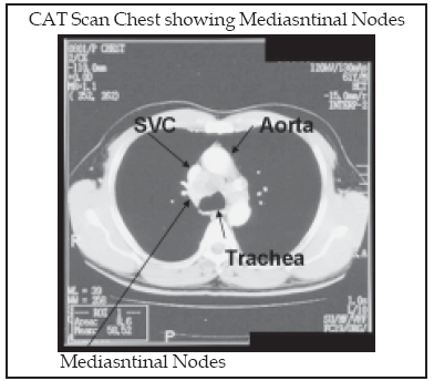
Since these lymph nodes are surrounded by great vessels and trachea (Fig-1), they are mostly inaccessible through the CT-guided approach. Even if CT biopsy could be performed, the biopsy specimens are very small and it is hard to diagnose lymphoproliferative malignancies.
3 Bronchoscopy & Trans-esophageal biopsies4 of these lymph nodes are achievable but again the diagnosis of lymphoma cannot be achieved due to small specimen. Surgical biopsies are the best ways to biopsy the mediastinal nodes. There are three ways to do this: mediastinoscopy via cervical neck incision, mediastinotomy via anterior chest incision in the second or third intercostal space, or via video-assisted thoracoscopy (VATS).5Although mediastinoscopy provides us with an approach to biopsy these nodes,
4 there are drawbacks to this procedure, which include, the small size of the biopsy specimens, inability to visualize the entire chest cavity and the propensity to form a big scar in the neck, which is a constant source of embarrassment especially for young girls in this part of the world. On the other hand, video-assisted thoracoscopy requires only a 2 cm incision in the chest at the fifth intercostal space. With this approach, one is able not only to obtain bigger samples and better visualization of the pleural cavity, but from the cosmetic stand point the scar is also not visible to other people.5The patient is placed in the lateral decubitus position and the procedure is performed under general anesthesia with a regular single lumen endotracheal tube. One or two <2cm incisions
are made in the fifth intercostal space just posterior to the anterior axillary line. (Fig-2). A 0° scope is used which is the same as a laparoscope.

Looking at the monitor, the targeted nodal station in the mediastinum is approached. The parietal pleura is lifted up using a grasper and cauterized with care so as not to injure the superior vena cava, phrenic nerve, aorta or pulmonary artery. The nodes are mobilized with blunt dissection via endo forceps and then multiple biopsies are taken under direct visualization on the TV monitor. The same principles are followed for nodal biopsies in the anterior or posterior mediastinal compartment with the only exceptions being the site of incision on the chest. The main contraindication to VATS includes the absence of a pleural space (i.e. previous thoracotomy) or inability to tolerate general anesthesia. The procedure requires only a 24-hour hospital stay and carries almost no risk.
To conclude, mediastinal adenopathy remains a Dilemma in TB endemic South Asian countries. However mediastinoscopy and especially video-assisted thoracoscopy (VATS) has changed the way mediastinal adenopathy is diagnosed and managed in this part of the world. These are the effective tools for obtaining a histological diagnosis of the disease, thus helping in accurate treatment of the patient by the pulmonologist or physician.
REFERENCES
1. Olak J. Benign Lymph Node Disease Involving the Mediastinum. In: Shields TW. General Thoracic Surgery 6th ed. Lippincott Williams & Wilkins Co; 2005;2676-2681.
2. World Health Organization- Estimated incidence, prevalence and TB mortality, 2004 In South East Asia.
3. Gossot D, Girard P, Eric de Kerviler, Brice P, Rain JD, Leblanc T, Grunenwald D. Thoracoscopy or CT-Guided Biopsy for Residual Intrathoracic Masses After Treatment of Lymphoma Chest 2001;120(1):289-294.
4. Yasufuku K, Chiyo M, Sekine Y, Chhajed PN, Shibuya K, Iizasa T, et al. Real-time Endobronchial Ultrasound-Guided Transbronchial Needle Aspiration of Mediastinal and Hilar Lymph Nodes Chest 2004;126(1):122-128.
5. Rendina EA, Venuta F, DeGiacomo T, Ciriaco PP, Pescarmona EO, Francioni F, et al. Comparative merits of thoracoscopy, mediastinoscopy, and mediastinotomy for mediastinal biopsy. Ann Thorac Surg 1994;57:992-995.
HOME | SEARCH | CURRENT ISSUE | PAST ISSUES
Professional
Medical Publications
Room No. 522, 5th Floor, Panorama Centre
Building No. 2, P.O. Box 8766, Saddar, Karachi - Pakistan.
Phones : 5688791, 5689285 Fax : 5689860
pjms@pjms.com.pk