|
|
||||
|
Published by : PROFESSIONAL MEDICAL PUBLICATIONS |
||||
|
ISSN 1681-715X |
||||
|
||||
|
- |
||||
|
ORIGINAL ARTICLE |
||||
|
- |
||||
|
Volume 23 |
October - December 2007 (Part-II) |
Number 6 |
||
|
|
||||
|
|
||||
|
|
||||
|
Published by : PROFESSIONAL MEDICAL PUBLICATIONS |
||||
|
ISSN 1681-715X |
||||
|
||||
|
- |
||||
|
ORIGINAL ARTICLE |
||||
|
- |
||||
|
Volume 23 |
October - December 2007 (Part-II) |
Number 6 |
||
|
|
||||
|
|
||||
Identification of aspergillus species
using morphological characteristics
K Diba1, Kordbacheh P2, Mirhendi SH3, S Rezaie4, M Mahmoudi5
ABSTRACT
Objectives: Although molecular methods continue to improve and become more rapidly available, microscopy and culture remain commonly used and essential tools for identification of Aspergillus spp. In this study we emphasize on morphological methods including; macroscopic and microscopic characteristics for identification of Aspergillus species isolated from environmental and clinical specimens.
Methodology: We used four differential media: czapek dox agar (CZ), czapek yeast agar (CYA), malt extract agar (MEA), and czapek yeast 20% sucrose agar. Morphological features of colonies on above culture media as well as microscopically characteristics for the major strains were studied and then compared with those of standard Aspergillus strains. Our major subjects were Iranian Aspergillus strains isolated from clinical and environmental specimens. Standard Aspergillus strains for study development included; A. fumigatus, (JCM 10253), A. flavus (JCM 2061), A. niger (JCM 10254), A. nidulans (JCM 02728), A. tereus (JCM 10227). Morphological features of Aspergillus cultures were studied, the major and remarkable macroscopic features in species identification were the colony diameter, color (conidia and reverse), exudates and colony texture. Microscopic characteristics for the identification were conidial heads, stipes, color and length vesicles shape and seriation, metula covering, conidia size, shape and roughness also colony features including diameter after 7 days, color of conidia, mycelia, exudates and reverse, colony texture and shape. Finally we compared the morphological characteristics of tested Aspergillus isolates with those of the standard species
Results: Aspergillus isolates were identified in the level of species using the differential culture media. A total of 205 Aspergillus isolates studied included: 153(75%) environmental Aspergilli and 52 (25%) clinical isolates. Within 11 Aspergillus species identified, A.flavus (55%), A.niger (31.7%) and A. fumigatus (8.7%) were the most common Aspergillus isolates from all of the specimens.
Conclusion: In our view morphological method using the differential media is the most reliable and sensitive assay to identify more medically important Aspergillus species.
KEY WORDS: Aspergillus, Identification, Macroscopic feature, Microscopic characteristics.
Pak J Med Sci October - December 2007 (Part-II) Vol. 23 No. 6 867-872
1. K Diba,
2. P. Kordbacheh,
3. SH.Mirhendi,
4. S. Rezaie,
1-4: Department of Parasitology and Mycology,
5. M Mahmoudi, Dept. of Epidemiology and Biostatistics,
1-5: School of Public Health and Institute of
Public Health Research,
Tehran University of Medical Sciences, Tehran – Iran.
Correspondence
Dr. K Diba,
E-mail: kdiba@razi.tums.ac.ir
* Received for Publication: April 13, 2007
* Revision Received: April 22, 2007
* 2nd Revision Received: September 29, 2007
* Final Revision Accepted: October 25, 2007
INTRODUCTION
Aspergillus species are frequent causes of invasive fungal infections in immunocompromised patients; they are also associated with allergic bronchopulmonary diseases, mycotic keratitis, otomycosis and nasal sinusitis.
1 At least 30 Aspergillus species have been associated with human diseases, and A. fumigatus remains the most frequent cause of invasive Aspergillosis (IA) but recently in some institutions, A. tereus is becoming more common and it is less susceptible to amphotericine B.2 A. nidulans has also been reported to be less susceptible to this drug in comparison with A. fumigatus. A. ustus as a rare cause of invasive disease has been, reported to be resistant to amphotericine B while remaining susceptible to itraconazole. A. ustus has also been reported to be less susceptible to voriconazole.2 Therefore accurate identification of species is important for the management of IA as well as for surveillance and other epidemiological purposes.Generally identification of the Aspergillus species is based on the morphological characteristics of the colony and microscopic examinations.
1 Although molecular methods continue to improve and become more rapidly available, microscopy and culture remain commonly used and essential tools for identification of Aspergillus spp. The 2003 American society for microbiology (ASM) survey documented that 89% of laboratories performing mycological examinations (morphology based), 16% of them use serologic tests and fewer than 5% use molecular tests for identification of microbial pathogens.3 Isolation in culture and phenotypic identification of common clinical isolates of Aspergillus spp. is usually quick and easy. However, culture is often described as slow, perhaps creating misconception about its value for the detection of aspergilli. A. fumigatus is a rapid grower.4Given the continued reliance on microscopy and culture, the diagnostic value of these methods must be improved by procedural changes and adequate training of laboratory personnel. The use of potato dextrose, potato flake, malt extract, inhibitory mould agar, or similar sporolation agars as primary isolation media for Aspergillus app. may speed growth rate and the production of conidia. The addition of antibacterial agents to isolation media helps reduce time to identification by inhibiting bacterial overgrowth and reducing the need for subculture. The initial incubation of fungal media at 35-37şC instead of, or in addition to 30şC may speed the growth of some aspergilli.
5In this study we emphasize on morphological methods including macroscopic features of colonies and microscopic characteristics for identification of Aspergillus species isolated from environmental and clinical specimens. We used four differential culture media that enabled us to discriminate most of medically important Aspergillus species. As far as we know this is the first study of its kind for identification of Aspergillus species in Iran.
METHODOLOGY
Microorganisms: Standard Aspergillus strains for study development included; A. fumigatus, (JCM 10253), A. flavus (JCM 2061), A. niger (JCM 10254), A. nidulans (JCM 02728) and A. tereus (JCM 10227) that was provided from the Japanese Microbiologic Collection (JCM). Our major subjects were Iranian Aspergillus strains isolated from clinical and environmental specimens. The clinical specimens were obtained from laboratory of medical mycology, Institute of Public Health Research, Tehran University of Medical Sciences. Environmental specimens included outdoor and indoor samples obtained from four educational hospitals in Tehran, Esfehan and Kermanshah. Surface sampling was done by wiping moistened sterile swabs over a surface measured approximately 20cm˛ and air samples were achieved by replacing the media plates in the exposure of air flow on different sites at above mentioned hospitals.
Culture and identification:
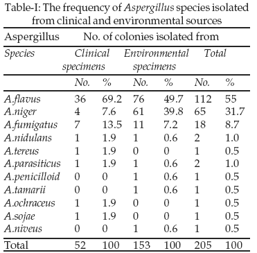
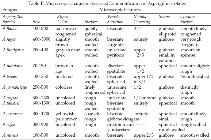
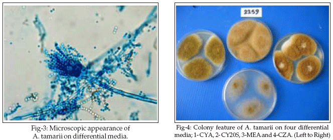
Aspergillus isolates were identified in the level of genus on Sabouraud Glucose Agar 4% (SGA4%). To improve the sensitivity and specificity of routine culture approach for identification of Aspergilli in the level of species, we used four differential media including, czapek dox agar ( CZ) {czapek concentration 10.0ml, K2HPO4 1.0gr, sucrose 30.0gr, agar 17.5gr, distilled water (DW) 1.0 lit (Maren A.Klich CBS-2002)}, czapek yeast agar (CYA) {czapek concentration 10.0ml, K2HPO4 1.0gr, powdered yeast extract 5.0gr, sucrose 30.0 gr, Agar 15.0gr and DW 1.0 lit., malt extract agar (MEA) {powdered malt extract 20.0gr, Peptone 10.0gr, Glucose 20.0gr, Agar 20.0gr, DW 1.0 lit} and czapek yeast 20% sucrose agar{ czapek concentration 10.0 ml, K2HPO4 1.0gr, powdered yeast extract 5.0gr, sucrose 200.0gr, agar 15.0gr, DW 1.0 lit}. Morphological features of Aspergillus cultures were studied, the major and remarkable macroscopic features in species identification were the colony diameter, color (conidia and reverse), exudates and colony texture. We used Riddle’s classic slide culture method6 for microscopic study of standard strains and most of our isolates. A quick method was used in some cases that was, simply to push an 18 × 18mm cover slip at 45 degree angle into the culture agar media. When the mould sporolated the cover slip was carefully withdrawn from the agar and mounted in a drop of lacto-fuchsine on a microscope slide. Another drop placed on top of the small cover slip before completing the assembly with a 22X 22mm cover slip. Microscopic characteristics for the identification were conidial heads, stipes, color and length vesicles shape and seriation, metula covering, conidia size, shape and roughness also colony features including diameter after 7 days, color of conidia, mycelia, exudates and reverse, colony texture and shape. As a final we compared the morphological characteristics of tested Aspergillus isolates with those of the standard species.RESULTS
During a period of 18-months, 205 isolates of Aspergillus species were obtained from the clinical and environmental specimens; The outdoor samples were collected from the air and surfaces of yard and garden of four Iranian teaching hospitals in Tehran, Esfehan and Kermanshah and indoor samples were collected from the air, floor, wall, beds, trolleys, air condition and the other surfaces. A total of 275 Aspergillus isolates were obtained which included: 153(75%) environmental Aspergilus isolates and the rest were 52 (25%) clinical isolates (Table-I). Clinical isolates were obtained from: 27 nail scrapping, 8 sinus discharge, 9 ear exudates, 3 sputum specimens and one biopsy material.
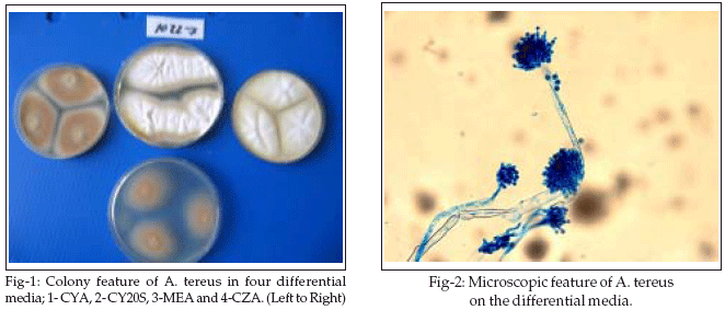
Hospital indoor Aspergillus isolates included: surgery wards, Delivery Rooms, operating rooms, ICUs and new borne wards; 24, 16, 12, 3, and 2 isolates respectively). Hospitals out door isolates (161) were obtained from the air and surfaces of the yard and garden by using four differential culture media; CZ, CYA, CYA20%S and MEA. We identified 11 Aspergillus species (Table-I). We also studied the five standard Aspergillus strains by using the above mentioned culture media. Morphological characteristics used for identification of Aspergillus species are showed in Table-II.
Our results show that in this study A. flavus (55%), A.niger (31.7%) and A. fumigatus (8.7%) were the most common isolated species from clinical an environmental specimens. Environmental isolated Aspergillus species included A. flavus (47.7%) from: indoor (29.8%) and outdoor (58.8%) isolates, A.niger totally 39% from indoor and outdoor isolates (56.1%) and (28.7%) respectively, followed by A. fumigatus 7.9% from indoor and outdoor isolates (14%) and (4.2%) respectively. Clinical isolates included; A. flavus (69.5%), A. fumigatus (13.5%), A. niger (7.6%) (Table-I).
DISCUSSION
More than thirty Aspergillus species have been associated with human diseases.
7 A. fumigatus remains the most frequent cause of invasive Aspergillosis (IA) but recently in some institutions, A. tereus is becoming more common and it is less susceptible to amphotericine B.2 A. nidulans has also been reported to be less susceptible to this drug in comparison with A. fumigatus. A.ustus as a rare cause of invasive disease has been, reported to be resistant to amphotericine B while remaining susceptible to itraconazole. A.ustus has also been reported to be less susceptible to voriconazole.2 As regards to different susceptibility of Aspergillus species to antifungal drugs accurate identification of species is important for management of invasive infections as well as for epidemiological purposes. In this study 11 Aspergillus species were identified by using the differential media, we demonstrated that use of four differential media including; CYA, CYA20S, MEA and CZ, was a simple and reliable method for identification of Aspergillus species. Although some studies with similar design was reported by klich,4 Mc Clenny1 and Luke Curtis,8 but this is the first Iranian study of its kind for identification of Aspergillus species. A recent similar douche study identified seven species of Aspergillus isolated from the water, air and surface samples.9 Another recent Spanish study identified Aspergillus spp. isolated from damp walls paper and the other surfaces.10
The result of our study showed that 11 species of Aspergillus in most common for A. flavus, A. niger and A. fumigatus respectively isolated from clinical and environmental (indoor and outdoor) specimens. The rare isolated Aspergillus species were A. parasiticus, A.sojae and A.niveus. A. flavus was the most common isolated species from the clinical specimens which included included nail scrpping, sputum, sinus discharge, ear exudates and biopsy materials. Our study showed that recovery of Aspergillus species from the nail scrapping was higher than other clinical specimens. Our findings from environmental specimens were in agreement with other studies that generally showed the higher amount of air borne spores of Aspergillus spp. in hospital outdoors. Therefore spores of Aspergillus could be transported by visitors, health care workers and medical devices from outside into wards. As the report by Anissie9 discussed the incidence of mold infections are increasing despite the wide spread use of air filtration systems that suggests other sources for distribution of Aspergillus spores. Study of Anissie identified A. niger (16%), A.fumigatus (17%) A. nidulans (7%), A.tereus (5%), A.flavus (3%), A.versicolor (1.5%) and A. clavatus (0.7%). In that study molds that were recovered on the interior surfaces included; A. niger (50%), A. fumigatus (19%), A.tereus (14%), A. flavus (9%), A.nidulans (7%) and A. sydowi (1%). Our study results showed a difference in the association of A. niger and A. flavus with air and surfaces swabs. Moreover our findings of A. flavus are not in agreement with Eliass11 report because of high frequency isolation after A. niger.
On the other hand our study data show that A.flavus is the most frequent species within all of Aspergillus species isolated from clinical specimens that followed by A.niger and A. fumigatus. Our findings are in agreement with data of a mycological research in Saudi Arabia12 on the nasal Aspergillus flora which were included A.flavus, A.niger & A.fumigatus respectively but that is in contrast with German study on outbreak of nosocomial Aspergillosis.13 Also in the study of Eliass11 it was observed that A.fumigatus was the predominant species isolated from blood and respiratory specimens, A.flavus was predominantly isolated from nasal polyps whereas A.niger predominated in nail specimens. Fridkin14 reported most frequent Aspergillus Species isolated from the air of a hematological hospital for A. fumigatus and A. flavus with a low different in frequency; 44% and 42% respectively.
Our study showed that single medium SGA 4% is only useful for identification of Aspergilli in genus level. In this study we used morphological method with four differential culture media identification of at least 11 medically important Aspergillus species. Using this method, five standard strains were identified successfully. Although isolation in culture and phenotypic identification of common clinical isolates of Aspergillus spp. is usually quick and easy. However, culture is often described as slow, perhaps creating misconception about its value for the detection of aspergilli. A. fumigatus is a rapid grower. The typical velutinous, gray-blue-green colonies uniseriate conidial heads develop within 24-48 h on both fungal media and the sheep blood agar commonly used for bacterial culture. Other aspergilli associated with invasive aspergillosis, specifically, A. flavus, A. niger, A. nidulans and A. terreus have growth rates similar to that of A. fumigatus when colonies were measured on malt extract agar and czapek yeast agar after incubation for seven days at both 25şC and 37şC.4 The use of potato dextrose, potato flake, malt extract, inhibitory mould agar, or similar sporolation agars as primary isolation media for Aspergillus app. may accelerate growth rate and the production of conidia5,15 and the identification of aspergilli based on morphological methods requires adequate growth for evaluation of colony characteristics and microscopic features. A culture time of 5 days or more is generally required for identification of anamorphic forms of Aspergillus. In our study using four differential media including CZ, CYA, CY20S and MEA with macroscopic and microscopic characteristics of fungal growth on this culture media enabled us to discriminate 11 medically important Aspergillus species. We recommend this sensitive and reliable method for identification of Aspergillus species isolated from some sources. Further studies would be helpful in clarifying the media and conditions most effective for the recovery and identification of clinically important aspergilli.
ACKNOWLEDGMENTS
The authors thank Professor K. Makimura (Institute of Medical Mycology, Teikyo University, Tokyo, Japan) for the standard Aspergillus species and thank Kh Omidi and A. Norouzinejad (Mycology lab of Institute of Health Research of Tehran University) for their assistance. This work was supported by the Vice Chancellor of Research, School of Public Health and Institute of Health Research, Tehran University of Medical Sciences.
REFERENCES
1. McClenny N. Laboratory detection and identification of Aspergillus species by microscopic observation and culture. J Md Mycol 2005;1:S125-S128.
2. Moody SF. Restriction Enzyme Analysis of mitochondrial DNA of the Aspergillus flavus group; A. flavus, A. parasiticus and A. Nomius. J Applied Environmental Microbiology 1990;2441-52.
3. Warris A, Voss A, Verweij PE. Hospital sources of Aspergillus species: new routes of transmission. J Rev Microbiology 2001;18:156-62.
4. Klich M. Identification of Common Aspergillus Species. Utrech. The Netherlands: Centraalbureeau voor Schimmelculture, 2002.
5. Tarrand JJ, Kontoiannis D, Han X. Culture incubation conditions affect the growth of Aspergillus spp. in an invitro model system of issue phase fungal growth. In 42nd ICAAC Abstracts, San Diego, California, September 27-30,2002. Washington, DC: ASM press, 2002;909.
6. Riddle RW. Permanent stained mycological preparation obtained by slide culture. Mycologia 1950;42:265-70.
7. De Aguirre L, Hurst SF, Choi JS. Rapid differentiation of Aspergillus Species from other medically important opportunistic molds and yeasts by PCR-Enzyme Immunoassay. J Clin Microbiol 2004;(42)8:3495-504.
8. Curtis L, Baker K. Aspergillus surveillance at a large tertiary – care hospital. J Hospital Infect 2005;59:188-96.
9. Anaissie EJ, Stratton SL. Dignani MC. Pathogenic molds including Aspergillus spp. in hospital water distribution systems. Rev Am S Hematol 2003;45:493-6.
10. Leenders AC, Belkum AV. Density and Molecular Epidemiology of Aspergillus in Air and relationship to outbreaks of Aspergillus infection. J Clin Microbiol 1999;1752-7.
11. Elliass D, Marriot D, Hajjeh RA, Warnock D, Meyers W. Surveillance of fungal infections. J Med Mycol 2000;38:173-82.
12. Saleh S. Al-Hedaithy. The yeast species causing fungemia at a university hospital in Riadh, SaudiArabia. J Mycoses 2002;46:293-8.
13. Nardoni S, Mancianti F, Sgorbini M, Taccini F. Identification and seasonal distribution of airborne fungi in three horse stables. J Mycopathologia 2005;160:29-34.
14. Fridkin SK, Jaris WR. Epidemiology of nosocomial fungal infections. J Clin Microbiol Rev 1996;499-511.
15. Centraalbureeau voor Schimmelculture (http://www.cbs.knaw.nl). Utrecht, The Netherlands: CBS; ©2004 (cited 2004 Aug 27). Aspergillus Reference Cultures. Available from: http://www.cbs.knaw.nl/icpa/index.htm
HOME | SEARCH | CURRENT ISSUE | PAST ISSUES
Professional
Medical Publications
Room No. 522, 5th Floor, Panorama Centre
Building No. 2, P.O. Box 8766, Saddar, Karachi - Pakistan.
Phones : 5688791, 5689285 Fax : 5689860
pjms@pjms.com.pk